Journal of Medical Cases, ISSN 1923-4155 print, 1923-4163 online, Open Access
Article copyright, the authors; Journal compilation copyright, J Med Cases and Elmer Press Inc
Journal website https://jmc.elmerpub.com

Case Report

Volume 17, Number 4, April 2026, pages 176-182


Successful Conservative Management of Complicated Brucella Endocarditis

Figures

↓  Figure 1. Admission brain computed tomography without contrast obtained on admission showing a well-defined hypodense lesion in the right (arrow), consistent with an acute embolic ischemic infarction (a). Cerebral angiography showed occlusion of the right middle cerebral artery (b).
Figure 1.
↓  Figure 2. Mid-esophageal view transesophageal echocardiogram (TEE) demonstrates: (a) pedunculated vegetation (lower arrow, measured 3 × 6 mm) on the bioprosthetic aortic valve cusp, and paravalvular aortic-root abscess (upper arrow), indicating advanced infective endocarditis. (b) Doppler echocardiogram shows left ventricular outflow tract (LVOT) fistula to the abscess cavity.
Figure 2.
↓  Figure 3. Transesophageal echocardiography (TEE) shows destructive prosthetic aortic valve endocarditis with peri-annular extension: (a) large mobile vegetation attached to the bioprosthetic aortic valve cusps, (b) extension of infection to the aortic annulus with paravalvular aortic root abscess formation, (c) destruction of the mitral–aortic intervalvular fibrosa (MAIVF) (arrow), and (d) a sinus of Valsalva pseudoaneurysm (arrow).
Figure 3.

Table

↓  Table 1. Clinical Timeline of Events for the Admitted Case Before and After BE Presentation
 
Timeline Clinical events and findings Management CRP (mg/L) Procalcitonin (ng/mL) Brucella serology titers Outcome
BE: Brucella endocarditis; CRP: C-reactive protein; CT: computed tomography; Echo: echocardiography; TMP-SMX: trimethoprim-sulfamethoxazole.
5 months prior Intermittent fever, lower back pain. Blood culture: Brucella melitensis Rifampicin + ciprofloxacin for 6 weeks. 6 - 1:320 Symptom resolution
2 months post-treatment (admission) Recurrent fever, malaise, 10 kg weight loss, followed by acute left-sided hemiparesis. Hospital admission, diagnostic workup. 12 0.20 1:40 → 1:160 
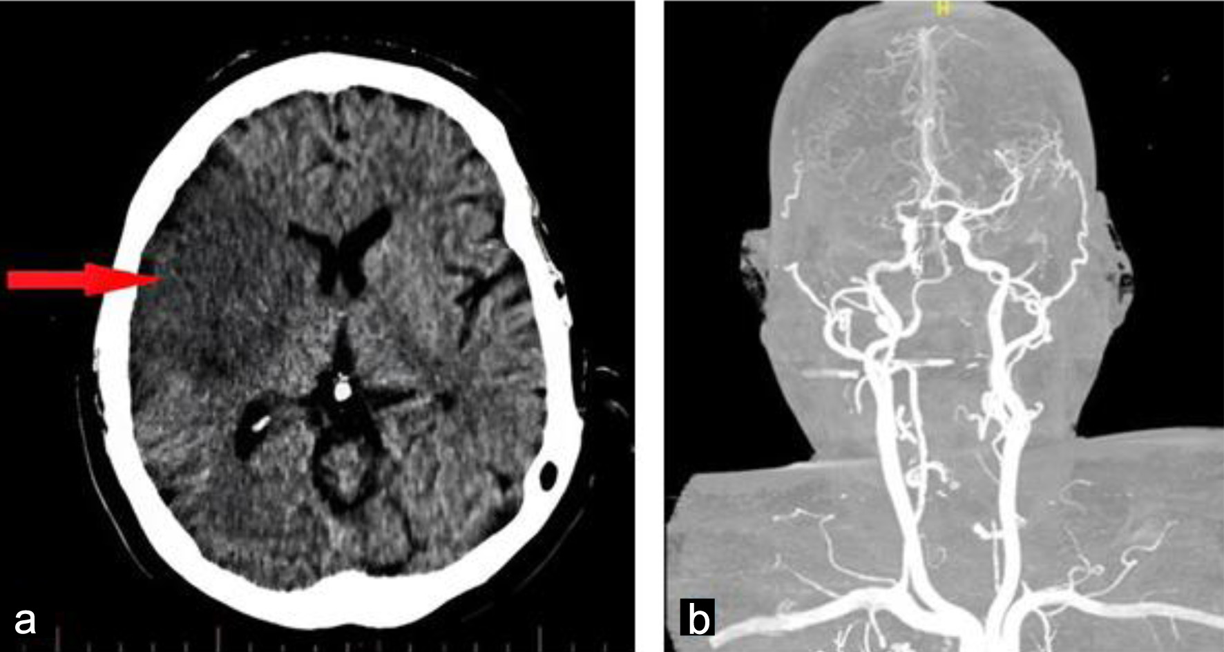
Embolic stroke confirmed on CT.
During inpatient phase (weeks 1–8) 
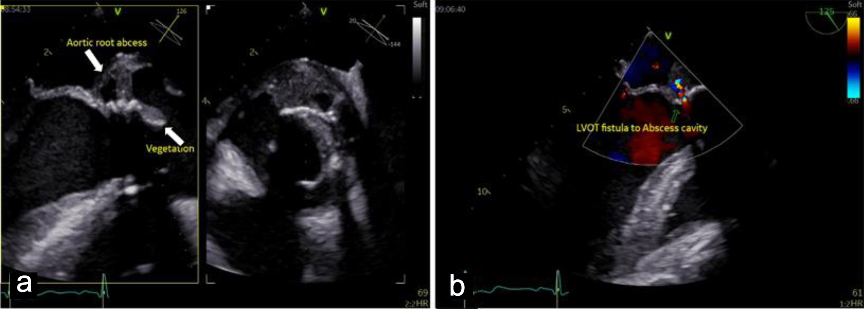
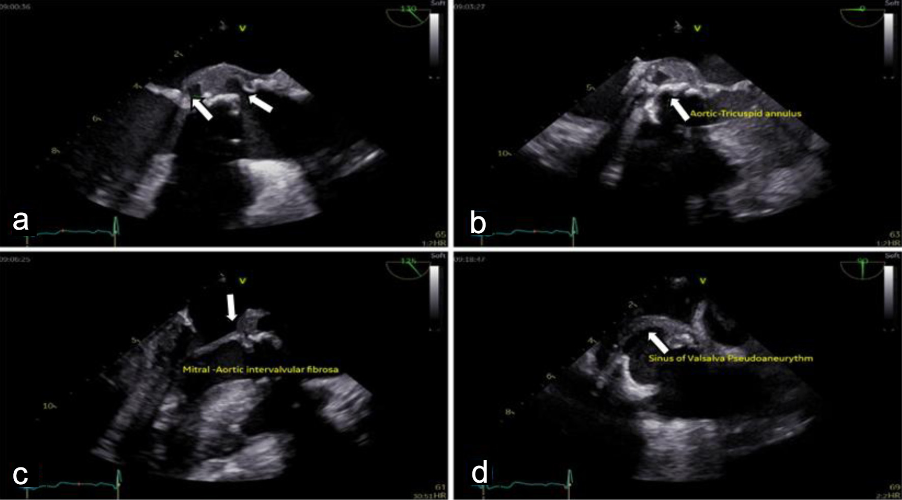
Diagnosis of BE with paravalvular abscess and spondylitis. Patient declined surgery. Initiated quadruple therapy: rifampicin, doxycycline, gentamicin, TMP-SMX. Inpatient monitoring. 14 → 10 → 3.37 0.01 → 0.03 1:320 Fever and back pain resolved. Neurological improvement. Echo: vegetation and abscess regressing.
Post-discharge (months 3–9) Outpatient follow-up. Gentamicin was discontinued after the initial phase. Continued oral therapy: rifampicin, doxycycline, TMP-SMX. Bi-weekly clinical and lab follow-up. 1.7 0.05 1:80 Clinically stable, afebrile. Independent in daily activities.
6-month follow-up Asymptomatic. No signs of heart failure or infection relapse. Completion of a prolonged antimicrobial course. Final echocardiogram. Normal Normal 1:40 (baseline) Complete resolution: No vegetation/abscess on echo. Preserved valve function. No relapse.