Journal of Medical Cases, ISSN 1923-4155 print, 1923-4163 online, Open Access
Article copyright, the authors; Journal compilation copyright, J Med Cases and Elmer Press Inc
Journal website https://jmc.elmerpub.com

Case Report

Volume 17, Number 9, September 2026, pages 439-448


Microscopic Polyangiitis With Pituitary Dysfunction and Spontaneous Renal Aneurysm Rupture

Figures

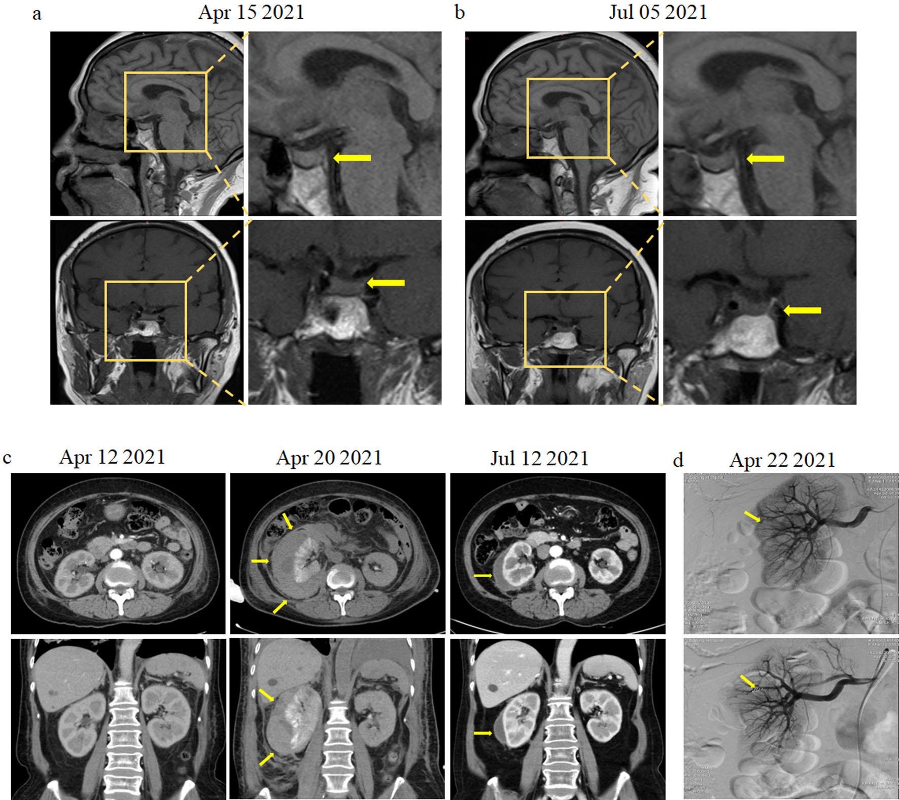
↓  Figure 1. Magnetic resonance imaging (MRI), computed tomography (CT), and arterial angiography images of the patient. (a) Sagittal and coronal pituitary T1-weighted MRI showing local signal enhancement (yellow arrow). (b) Sagittal and coronal pituitary T1-weighted MRI showing normal signal (yellow arrow). (c) Abdominal CT scan showing a large right-sided perinephric and intracapsular hematoma in the right kidney (yellow arrow). (d) Arterial angiography showing active bleeding from the superior segmental and interlobular renal artery of the right kidney. Disseminated arterial microaneurysms are indicated by a yellow arrow (upper panel). Highly selective arterial embolization of the interlobular artery of the right kidney using microcoils (lower panel).
Figure 1.
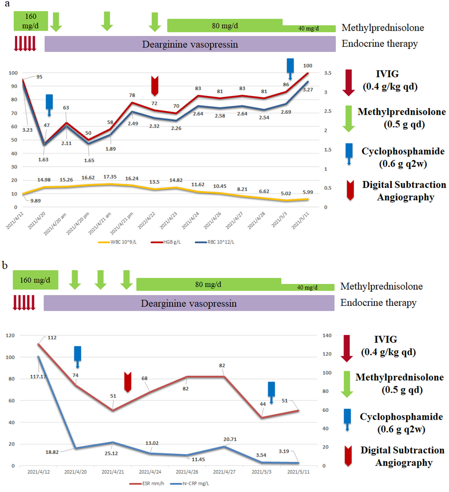
↓  Figure 2. Treatment strategies of this patient. (a) White blood cell (WBC), hemoglobin (HGB), and red blood cell (RBC) response in relation to the serial treatment. (b) Erythrocyte sedimentation rate (ESR) and C-reactive protein (CRP) levels in the serial treatment.
Figure 2.
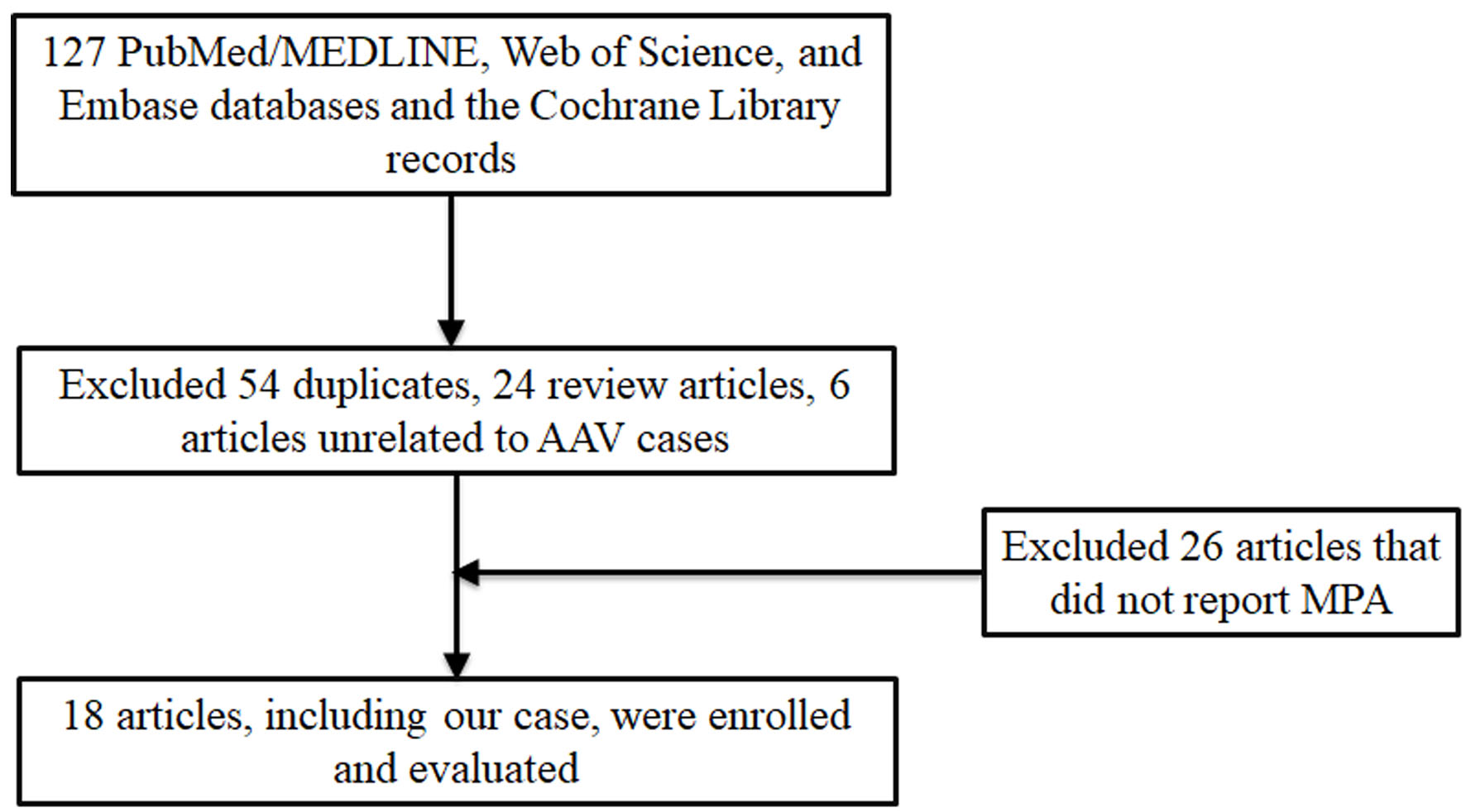
↓  Figure 3. Flow diagram of records in literature review process.
Figure 3.

Tables

↓  Table 1. Reported Cases of Pituitary Dysfunction in Patients With MPA
 
ItemsLi/2020 [6]Funauchi/2002 [7]Mercedes/2025 [8]This case
ANCA: antineutrophil cytoplasmic antibody; CTX: cyclophosphamide; F: female; HD-GC: high dose glucocorticoid; M: male; MPA: microscopic polyangiitis; MPO: myeloperoxidase; MRI: magnetic resonance imaging; PD-GC: pulse dose glucocorticoid; PET-CT: positron emission tomography-computed tomography.
Sex/ageM/61F/62F/41F/59
Auto-antibodyMPO-ANCAMPO-ANCAMPO-ANCAMPO-ANCA
Onset of pituitary injuryOnset of MPA2 months before MPA diagnose2 weeks before MPA diagnoseOnset of MPA
Symptoms of Pituitary injuryPolydipsia, polyuria, and erectile dysfunctionPolydipsia, polyuriaPersisted headache, sinus/facial discomfortPolydipsia, polyuria
Imaging findingsMRI (negative), PET-CT: increased linear radioactivity uptake in pituitary fossaMRI: swelling of the pituitary gland and loss of the high intensity signal of posterior lobeMRI: enlarged heterogeneous pituitary gland with infundibular thickeningMRI: enlargement of pituitary gland and local signal enhancement
TreatmentPD-GC and HD-GC + CTXPrednisolone for 4 weeksGC and rituximabPD-GC and HD-GC+CTX
Hormone replacementDesmopressinDesmopressinNoneDesmopressin
OutcomesPartial remissionRemissionRemissionRemission


↓  Table 2. Reported Cases of Renal Aneurysm in Patients With MPA
 
Case/yearSex/ageAuto-antibodyPresentationImaging examinationsInvolved arteryPerirenal hematomasTreatmentOutcomes
ANCA: antineutrophil cytoplasmic antibody; CT: computed tomography; CTA: computed tomography angiography; CTX: cyclophosphamide; F: female; HD: hemodialysis; IV MP: intravenous methylprednisolone; M: male; MPO: myeloperoxidase; NA: not available; PE: plasm exchange.
Inatsu/2002 [14]M/78MPO-ANCASlight fever and weight loss, RPGNArterial angiographyBilateral renal arteryNoSteroidsRecovered, HD
Tamei/2008 [11]M/55MPO-ANCASharp pain on the left side, shockAbdominal CT and arterial angiographyLeft renal, hepatic arteryYes, leftBlood transfusion, steroidsRecovered, HD
Dhaun/2013 [15]M/56MPO-ANCABilateral loin painCT and CTABilateral renal, hepatic, mesenteric arteryYes, bilateralSteroids + CTX, PERecovered
Jira/2013 [16]M/56MPO-ANCASevere right iliac fossa pain hemodynamic shockRenal arteriographyBilateral renal arteryYes, rightIV MP + CYC, oral steroidsRecovered
Nakashima/2015 [24]F/77MPO-ANCARPGN, both flank painAbdominal CT and ultrasonographyBilateral renal arteryYes, bilateralOral steroidsRecovered, HD
Ishiwatari/2018 [10]F/82MPO-ANCARPGN, decreased urinary volume, decreased hemoglobinAbdominal CTRight renal arteryYes, rightCoil embolization, IV MP, oral steroids mizoribineRecovered, HD
Yu/2018 [17]M/61MPO-ANCALeft lumbago, decreased hemoglobinContrast enhanced CTBilateral renal, hepatic, and mesenteric arteryYes, leftNephrectomy in emergencyNA
Zhang/2019 [18]F/57MPO-ANCASevere lumbago, acute renal failure, and hemorrhagic shockAbdominal CTBilateral renal arteryYes, bilateralIV MP, oral steroidsRecovered
Andrew/2020 [19]F/50MPO-ANCAAbdominal pain, decreased hemoglobinAbdominal CT, contrast enhanced CTBilateral renal, hepatic arteryYes, bilateralPercutaneous drainage of hematoma, IV MP and CYCNA
Nunes/2022 [20]M/52Negative (kidney biopsy)Severe abdominal pain and hemorrhagic shockCTALeft renal arteryYes, leftCoil embolization, IV MP + CYC, oral steroidsRecovered
Zhao/2023 [21]F/56MPO-ANCABack painAbdominal CTRight renal arteryYes, rightOral steroids + CTX, PERecovered
Tang/2024 [22]M/61MPO-ANCALow back pain, decreased hemoglobinRenal ultrasonography, arteriographyBilateral renal arteryYes, bilateralIV MP + CYC, PE and coil embolizationRecovered, HD
Jana/2025 [23]F/45MPO-ANCASevere abdominal pain, drop in hemoglobinAbdominal CT, MRI, and CTALeft renal arteryYes, leftIV MP + CYC, oral steroidsRecovered
This caseF/59MPO-ANCASharp pain in her right waist area, hypotension, decreased hemoglobinAbdominal CT, contrast enhanced CT, arteriographyRight renal arteryYes, rightCoil embolization, IV MP + CYC, oral steroidsRecovered