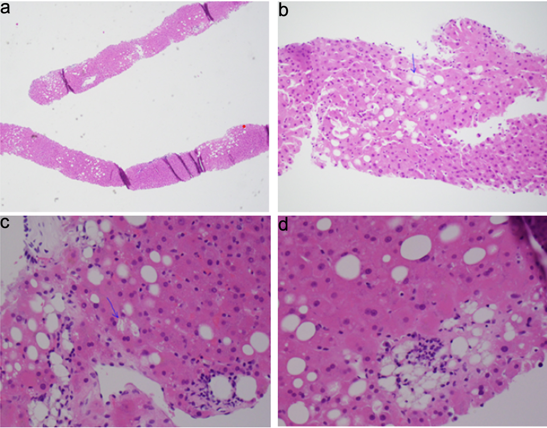
Journal of Medical Cases, ISSN 1923-4155 print, 1923-4163 online, Open Access
Article copyright, the authors; Journal compilation copyright, J Med Cases and Elmer Press Inc
Journal website https://jmc.elmerpub.com

Case Report

Volume 17, Number 3, March 2026, pages 121-127


Hemobilia as a Complication of Transjugular Liver Biopsy Causing Acute Pancreatitis and Obstructive Jaundice: A Case Report and Minireview

Figures

Figure 1.
Figure 1. Histopathology examination of hepatic parenchyma. (a) Steatosis, involving 10–15% of hepatocytes. (b, c) Ballooning of hepatocytes (arrows). (d) Steatosis and lobular inflammation.
Figure 2.
Figure 2. Magnetic resonance cholangiopancreatography. (a) Stricture (arrow) noted in common bile duct. (b) Dilated bile duct with distal stricture (arrow).
Figure 3.
Figure 3. Cholangiogram. (a) Fluroscopic image revealing wire cannulation of bile duct. (b) Cholangiogram revealing a dilated biliary tree with filing defects.
Figure 4.
Figure 4. Endoscopic retrograde cholangiopancreatography. (a) Endoscopic sphincterotomy followed by balloon sweep and extraction of blood clots. (b) Plastic bile duct and pancreatic duct stents were deployed to maintain biliary drainage and to prevent post-ERCP pancreatitis, respectively.