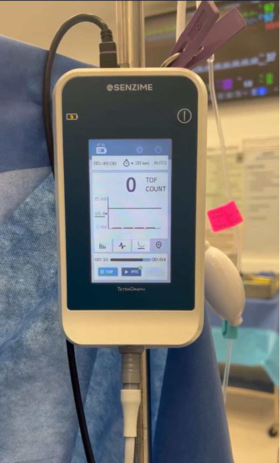
↓ Figure 1. TetraGraph™ electromyography
based quantitative TOF monitor (Senzime BV, Uppsala, Sweden). The displayed TOFC is 0/4. TOFC:
train-of-four count.
| Journal of Medical Cases, ISSN 1923-4155 print, 1923-4163 online, Open Access |
| Article copyright, the authors; Journal compilation copyright, J Med Cases and Elmer Press Inc |
| Journal website https://jmc.elmerpub.com |
Case Report
Volume 17, Number 4, April 2026, pages 163-169
Intraoperative Neuromuscular Monitoring: Electromyography Monitor Versus Peripheral Nerve Stimulator. Does the Face Lie?
Figures
Table
| Absolute time | Rocuronium bolus (mg/kg) | Rocuronium infusion rate (mg/kg/h) | EMG TOFC (twitches) | PNS assessment | Event |
|---|---|---|---|---|---|
| EMG: electromyography; PNS: peripheral nerve stimulator; TOFC: train-of-four count; TOFR: train-of-four ratio. | |||||
| 8:05 | — | — | — | — | Induction |
| 8:17 | 1.2 | — | Not assessed | Not assessed | |
| 8:19 | — | — | 0/4 | Not assessed | Endotracheal intubation |
| 8:52 | — | Start (0.6) | 1/4 | 1/4 | |
| 9:04 | 0.4 | 0.72 | 2/4 | 2/4 | |
| 9:55 | — | 0.6 | 0/4 | 0/4 | |
| 10:47 | — | 0.48 | 0/4 | 1/4 | |
| 11:07 | — | 0.48 | 0/4 | 1/4 | |
| 11:40 | — | 0.36 | 0/4 | 2/4 | |
| 12:08 | — | 0.36 | 1/4 | 4/4 | |
| 12:15 | — | 0.36 | 1/4 | 4/4 | Onset of spontaneous breathing |
| 12:29 | 0.6 | 0.48 | 1/4 | 4/4 | |
| 12:31 | — | 0.48 | 0/4 | 0/4 | |
| 12:52 | — | 0.48 | 0/4 | 1/4 | |
| 13:52 | — | 0.48 | 0/4 | 2/4 | |
| 14:01 | — | Stop | 0/4 | 2/4 | |
| 14:07 | — | — | 0/4 | 2/4 | Onset of spontaneous breathing |
| 14:08 | 0.2 | — | 0/4 | 2/4 | |
| 14:51 | — | — | 1/4 | 4/4 | Sugammadex administration |
| 15:08 | — | — | 4/4 (TOFR was ≥ 0.9) | Not assessed | Tracheal extubation |