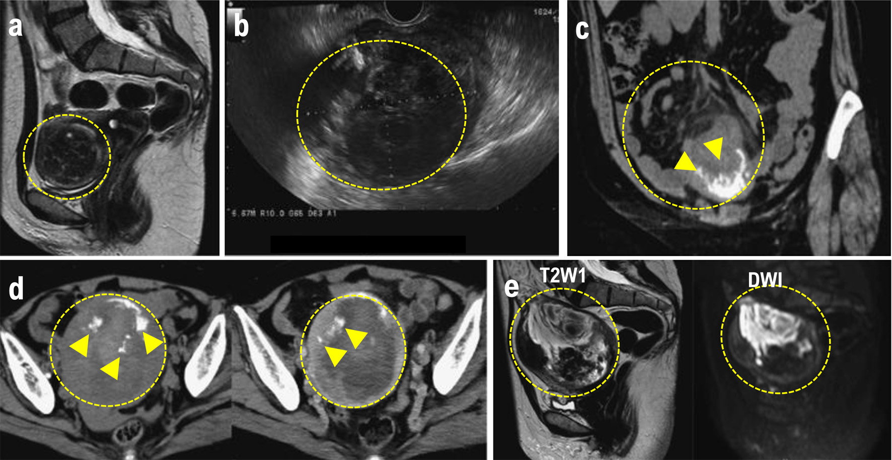
↓ Figure 1. (a) MRI performed at the initial
consultation. The mass exhibited a clearly demarcated low signal intensity on T2W1 and was diagnosed as
a uterine fibroid. (b) Transvaginal ultrasound after lower gastrointestinal endoscopy revealed a 90
× 69 mm mass in the uterine body, with a heterogeneous interior and irregular margins. (c) Plain
CT. Increased density was observed in the mesenteric fat tissue surrounding the mass. A mass with
marginal calcification was found on the anterior wall of the uterine body, protruding cephalad. (d)
Comparison of the initial CT (left) with a CT performed 8 days later for preoperative evaluation
(right). The uterine mass had shrunk from 10 × 11 cm (left panel) to 10 × 9.5 cm (right
panel), and the absorption value of the internal high-density area had decreased, suggesting absorption
of the hematoma. (e) MRI for further investigation of suspected uterine sarcoma. The mass showed
moderate signal intensity on T2W1 and high signal intensity on DWI, and heterogeneous signals were
present within it, suggesting hemorrhage and necrosis. The mass is indicated by the yellow dotted
circle. Heterologous components including osteosarcoma and chondrosarcoma are indicated by the white
arrowheads. CT: computed tomography; MRI: magnetic resonance imaging.