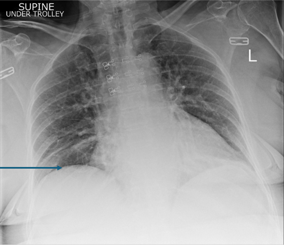
Journal of Medical Cases, ISSN 1923-4155 print, 1923-4163 online, Open Access
Article copyright, the authors; Journal compilation copyright, J Med Cases and Elmer Press Inc
Journal website https://jmc.elmerpub.com

Case Report

Volume 17, Number 4, April 2026, pages 153-156


Spontaneous Onset of Postoperative Chilaiditi Syndrome: A Case Report and Its Potential Other Association?

Figures

Figure 1.
Figure 1. Initial supine chest radiograph (CXR) revealed shallow lung expansion with elevated hemidiaphragms more in the right side. The arrow points towards elevated right hemidiaphragm. Image taken preoperatively.
Figure 2.
Figure 2. Anterior-posterior chest X-ray, 3 years prior to presentation showing normal chest X-ray and no gas loops in the hemidiaphragm.
Figure 3.
Figure 3. Anterior-posterior chest X-ray on the day of symptoms beginning showing bilateral gas filled loops beneath both hemidiaphragms more in the right side. The arrow points towards elevated right hemidiaphragm. Images taken day 1 postoperatively.
Figure 4.
Figure 4. Confirmed Chilaiditi, normal variant with normal appearances of the solid abdominal organs. No dominant lymphadenopathy. No free fluid. No pelvic mass. Images taken day 1 postoperatively.