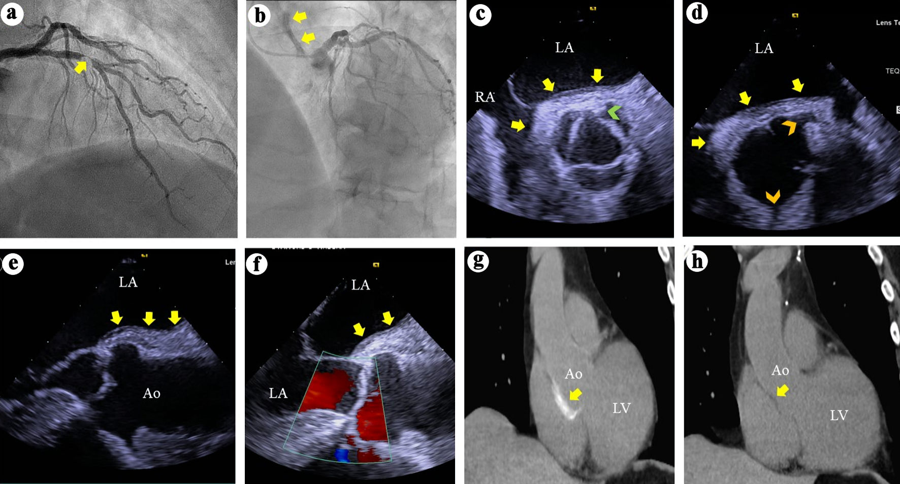
Journal of Medical Cases, ISSN 1923-4155 print, 1923-4163 online, Open Access
Article copyright, the authors; Journal compilation copyright, J Med Cases and Elmer Press Inc
Journal website https://jmc.elmerpub.com

Case Report

Volume 16, Number 12, December 2025, pages 499-503


Aortic Dissection During Diagnostic Coronary Angiography in a Patient With Acute Coronary Syndrome: The Role of Off-Pump Coronary Artery Bypass Grafting

Figure

Figure 1.
Figure 1. (a) Right anterior oblique view of the left coronary arteries angiography demonstrates a > 95% complex stenosis (arrow) of the left anterior descending coronary artery. (b) Left anterior and cranial view of the left coronary system demonstrates contrast hang-up or staining (arrows) on the aortic root and ascending aorta. (c) TEE short axis views at the aortic valve level during systole demonstrates soft tissue echo-reflectant thickening of the aortic root posterior wall with medial and lateral extensions (arrows) and a possible small intimal tear (green arrowhead). (d) TEE short-axis view of the aortic root during diastole demonstrates aortic wall thickening (arrows) and intact ostial left main and right coronary arteries (top and bottom arrowheads, respectively). (e, f) TEE long-axis views demonstrate soft tissue thickening of the posterior aortic root and ascending aortic wall (arrows) and intact aortic valve with no regurgitation. (g) Thoracic CT demonstrates contrast uptake at the aortic root and part of the ascending aorta (arrow). (h) Repeat thoracic CT 7 weeks later demonstrates resolution of the contrast uptake at the aortic root and ascending aorta. TEE: transesophageal echocardiography; CT: computed tomography; LA: left atrium; RA: right atrium; Ao: aorta; LV: left ventricle.

Table

Table 1. Iatrogenic Aortic Dissection (IAD): Insights From Three Retrospective Studies
 
AspectDunning et al, 2000 [1]Gomez-Moreno et al, 2006 [2]Nunez-Gil et al, 2015 [3]
ACS: acute coronary syndrome; PCI: percutaneous coronary intervention; CABG: coronary artery bypass grafting; RCA: right coronary artery.
Incidence0.02% (9/43,143 catheterizations)0.04% (18/40,900 catheterizations)0.07% (74/108,083 catheterizations)
Patient profileMostly ACS patients undergoing PCIPredominantly ACS cases during PCIPrimarily ACS cases during PCI
MechanismRCA dissection with retrograde extension into the aorta in all casesRetrograde coronary dissection into aorta in most casesSimilar: coronary dissection with retrograde aortic extension
Classification (Dunning)Class I: limited to coronary sinusNot specifically classifiedNot specifically classified
Class II: < 4 cm into ascending aorta
Class III: > 4 cm into ascending aorta
ManagementPCI (all 9 patients had stenting of the dissected RCA)Conservative (8 patients)Conservative (35 patients)
Surgery (especially for class III)PCI (10 patients)PCI (34 patients)
Surgery (3 patients: 2 aortic, 1 CABG)
None (2 patients, who died)
OutcomesSurgery in class III associated with worse prognosisDuring follow-up, 6 patients required not specified coronary or aortic surgical repairs.Overall mortality rate: 2.7%