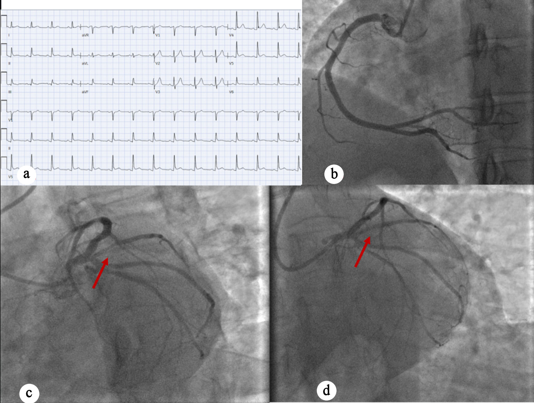
↓ Figure 1. (a) EKG showing sinus rhythm with no
acute ischemic changes. (b) Left anterior oblique caudal view showing right coronary artery. (c) Left
anterior oblique caudal view showing complete occlusion of left circumflex artery (red arrow). (d)
Post-PCI perfusion of the left circumflex artery (red arrow). EKG: electrocardiogram; PCI: percutaneous
coronary intervention.
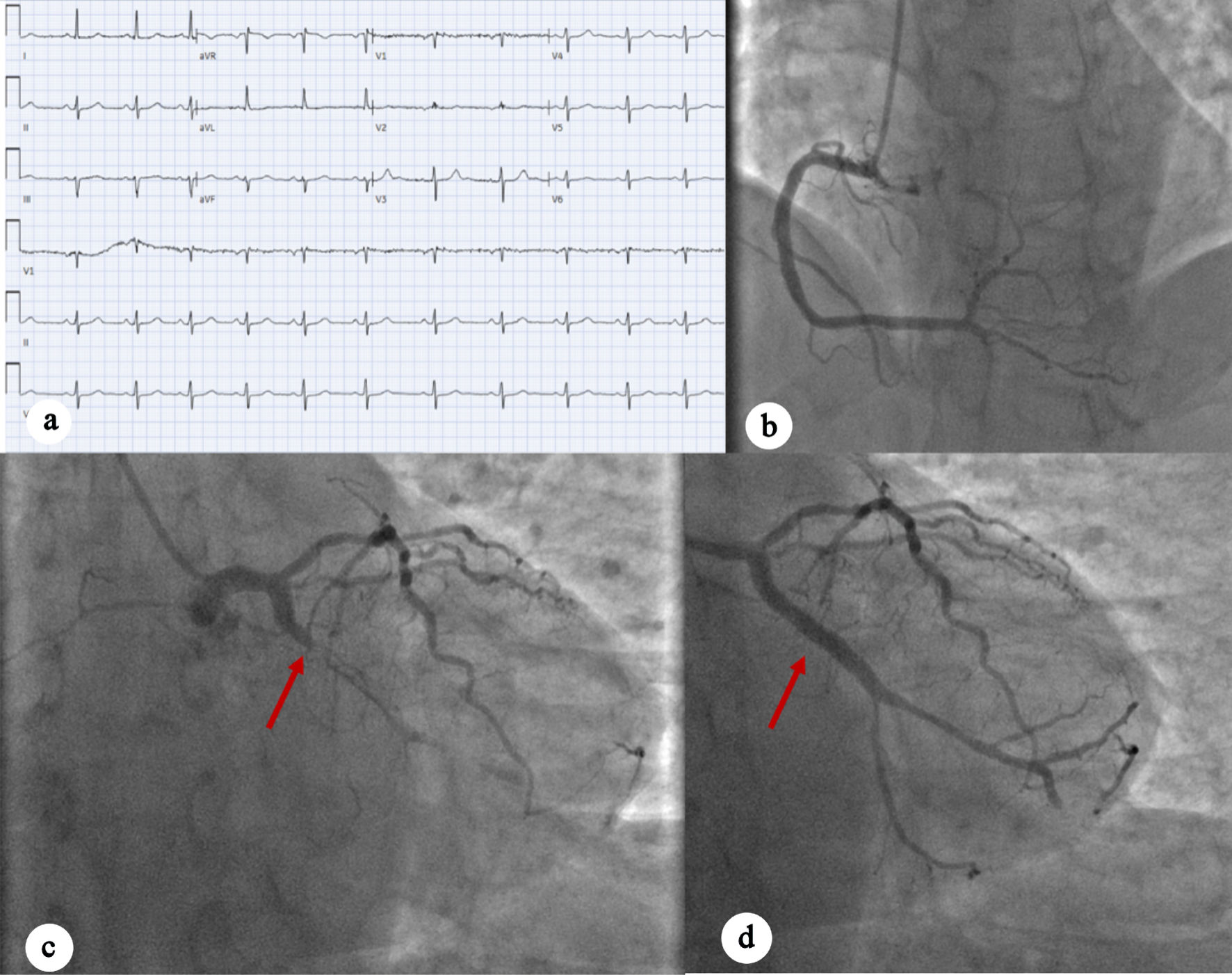
↓ Figure 2. (a) EKG showing normal sinus rhythm
with no acute ischemic changes. (b) Left anterior oblique caudal view showing early-mid-RCA with
moderate nonobstructive disease. (c) Left anterior oblique caudal view indicating complete occlusion of
OM 100% occlusion, as well as mid-LAD moderate disease (red arrow). (d) Left anterior oblique caudal
view showing post-PCI perfusion of OM1 (red arrow). EKG: electrocardiogram; RCA: right coronary artery;
LAD: left anterior descending artery; OM: obtuse marginal artery; PCI: percutaneous coronary
intervention.
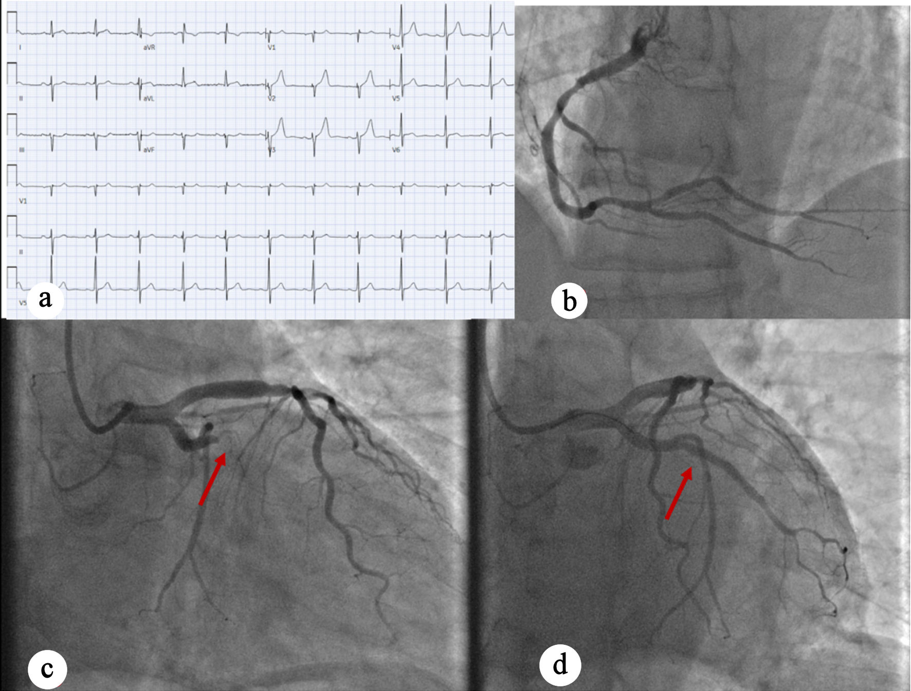
↓ Figure 3. (a) EKG showing normal sinus rhythm
with no ischemic changes. (b) Right anterior oblique caudal view showing normal RCA. (c) Coronary
angiography indicating 90% proximal ramus intermedius stenosis (red arrow). (d) Left anterior oblique
caudal view post-PCI perfusion of ramus intermedius (red arrow). EKG: electrocardiogram; RCA: right
coronary artery; LAD: left anterior descending artery; OM: obtuse marginal artery; PCI: percutaneous
coronary intervention.