Figures
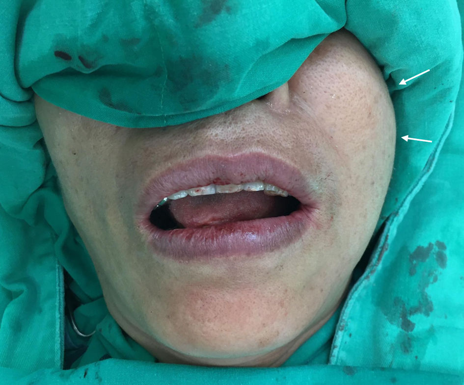
↓ Figure 1. Physical examination revealed a soft,
mobile, subcutaneous mass (about 3 cm) in the left buccal region (arrows).
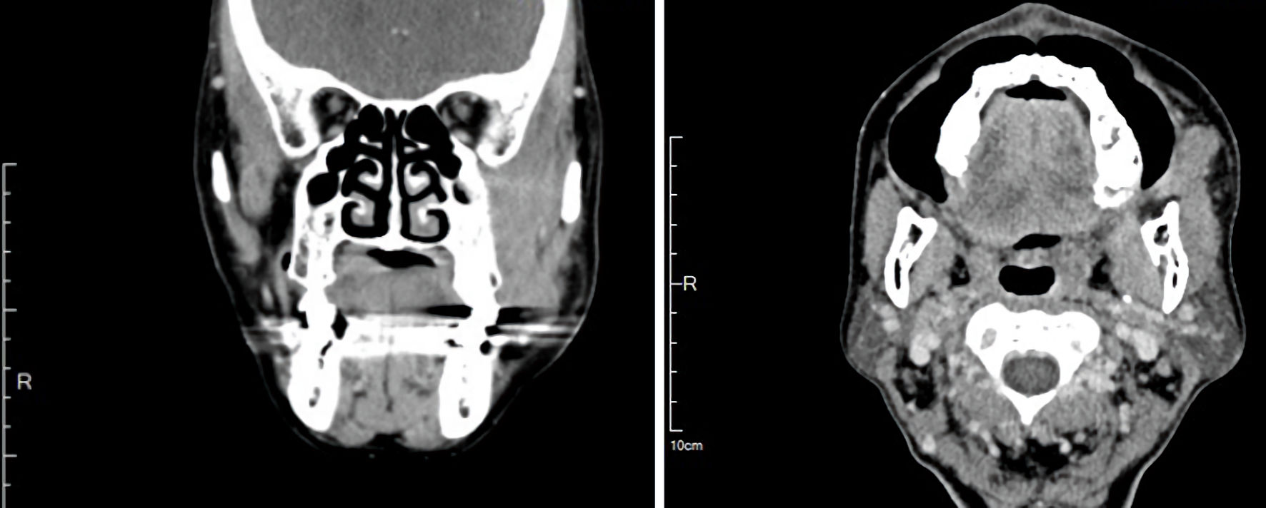
↓ Figure 2. Contrast-enhanced computed tomography
revealed an infiltrative soft-tissue mass measuring 3.1 × 1.5 × 3.5 cm, centered in the
masticator space with effacement of fat planes between the masseter, pterygoid, and temporalis
muscles.
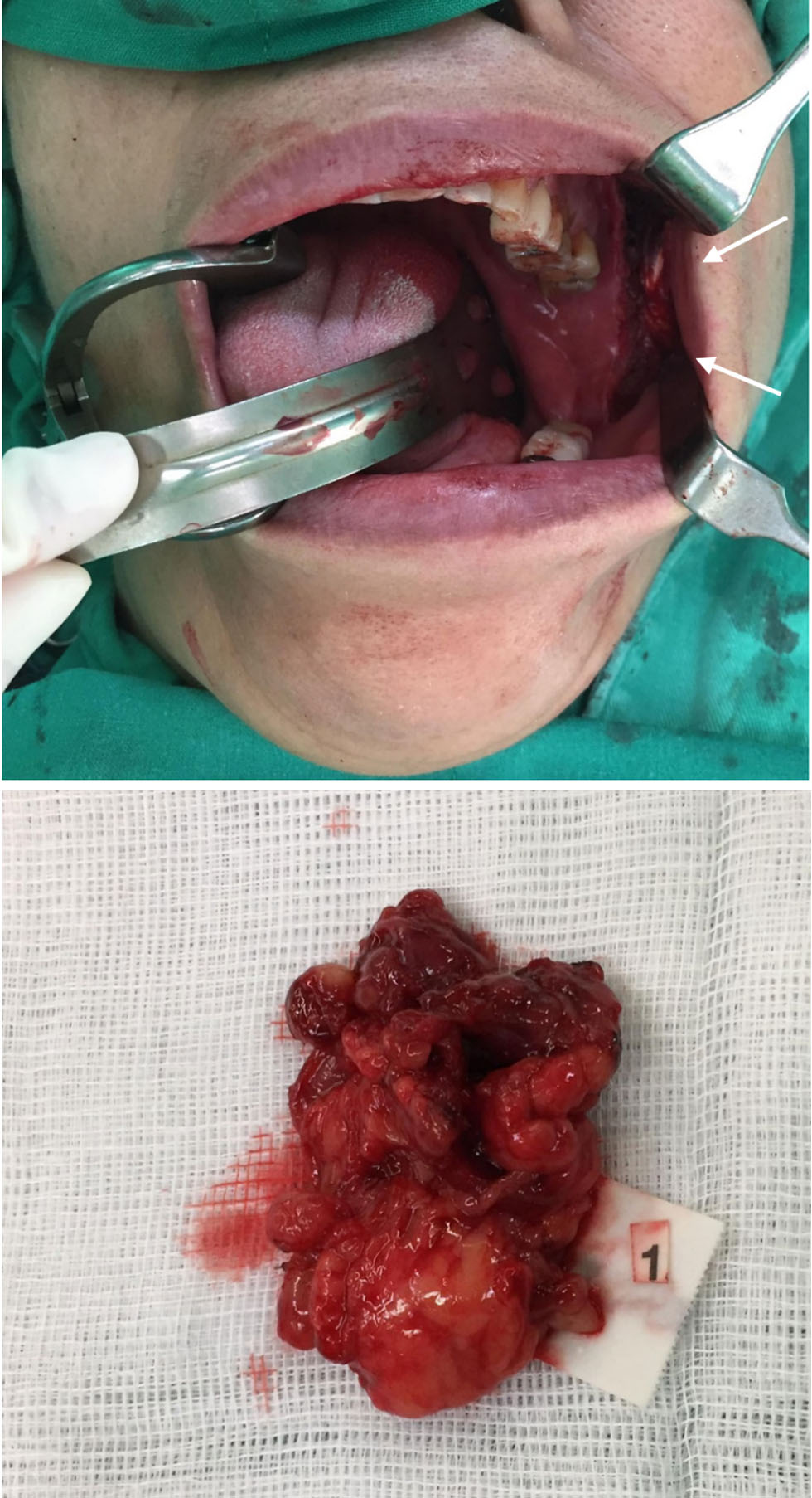
↓ Figure 3. An intraoral incisional biopsy under
general anesthesia with obtained specimen (arrows). The intraoral approach allowed direct access to the
bucco-masseteric space while avoiding external scarring.
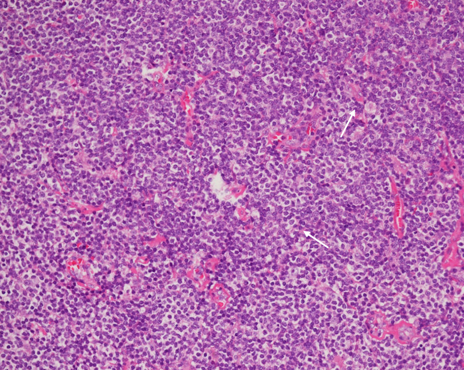
↓ Figure 4. H&E stain (× 40)
demonstrating a diffuse, vaguely nodular infiltrate, expanding the soft tissue and disrupting normal
architectural boundaries. Note the presence of mild plasmacytic differentiation (arrows). H&E:
hematoxylin and eosin.
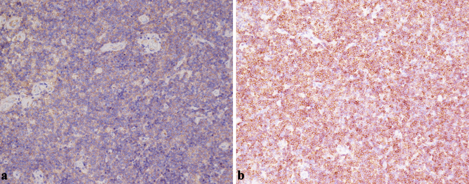
↓ Figure 5. (a) Representative
immunohistochemistry demonstrating diffuse CD20 positivity in the neoplastic B cells within the
masticator-space lesion (× 200). (b) Bcl-2 staining highlights strong cytoplasmic expression in the
small B cells, supporting a diagnosis of EMZL rather than reactive hyperplasia (× 200). EMZL:
extranodal marginal zone B-cell lymphoma.
↓ Figure 6. (a) CD3 negativity in the tumor cell
population confirms that the predominant infiltrate is of B-cell rather than T-cell lineage (×
200). (b) Lack of CD5 expression helps to exclude mantle cell lymphoma and most chronic lymphocytic
leukemia/small lymphocytic lymphoma (× 200). (c) The neoplastic lymphoid infiltrate is negative for
CD10, supporting a marginal zone origin and helping exclude follicular lymphoma (× 200). (d) The
neoplastic lymphoid population is negative for CD23 expression, helping distinguish this case from small
lymphocytic lymphoma (× 200). (e) CD43 negativity in the neoplastic B cells is consistent with an
EMZL phenotype and argues against other small B-cell lymphomas (× 200). (f) Bcl-6 negativity in the
neoplastic lymphoid cells (× 200). This lack of Bcl-6 expression supports a post-germinal center
origin and helps exclude follicular lymphoma. EMZL: extranodal marginal zone B-cell lymphoma.

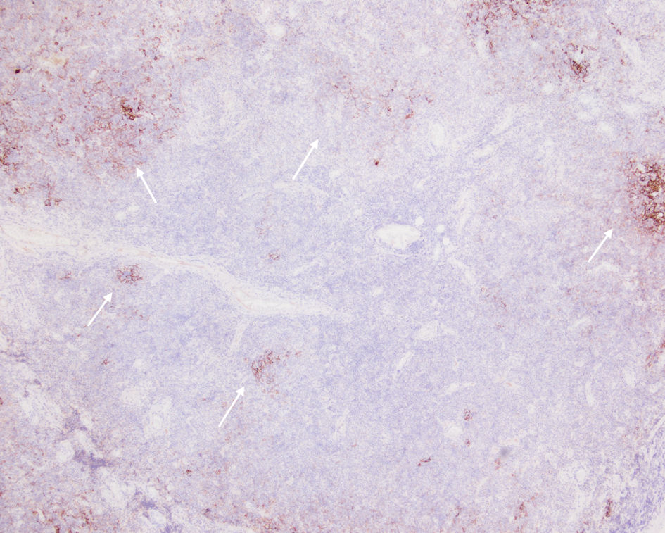
↓ Figure 7. CD21 outlining disrupted follicular
dendritic cell meshworks consistent with follicular colonization (arrows).