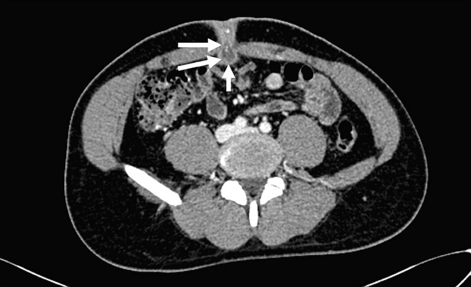
Journal of Medical Cases, ISSN 1923-4155 print, 1923-4163 online, Open Access
Article copyright, the authors; Journal compilation copyright, J Med Cases and Elmer Press Inc
Journal website https://jmc.elmerpub.com

Case Report

Volume 16, Number 11, November 2025, pages 456-460


A Rare Cause of Umbilical Discharge in a Healthy Adult: A Case Report of Patent Urachal Sinus in Primary Care

Figures

Figure 1.
Figure 1. (a) Pus discharge from the umbilicus during initial presentation at the primary care clinic. (b) Purulent discharge from the umbilicus upon gentle compression during initial presentation at the primary care clinic.
Figure 2.
Figure 2. Axial abdominal contrast-enhanced computed tomography image shows a blind ending, thick-walled, hypodense tubular collection (white arrows) extending posteriorly from the umbilicus into the abdominal cavity in keeping with an infected umbilical-urachal sinus.
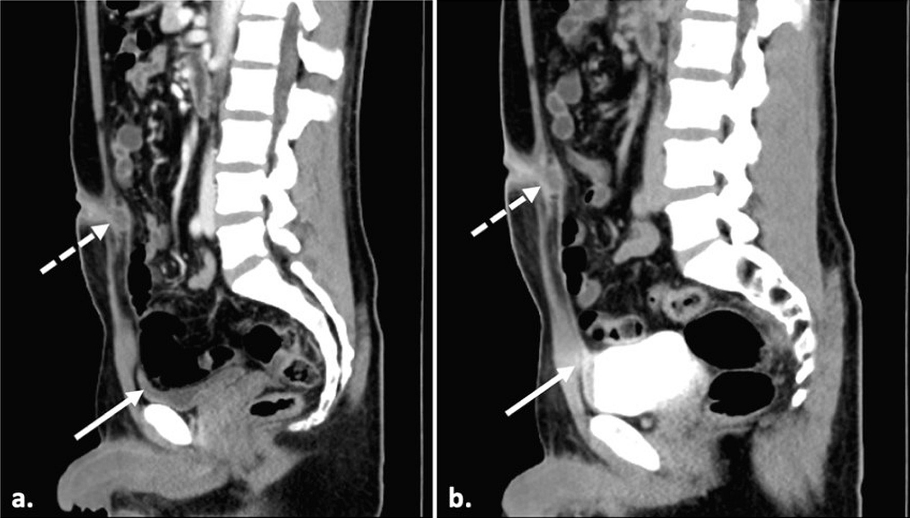
Figure 3.
Figure 3. Midsagittal CT scan images of (a) contrast-enhanced scan and (b) delayed images with a full bladder showing the partially filled vesicourachal diverticulum (solid arrow) and the infected umbilical-urachal sinus (dashed arrow). There is no evidence of communication of the umbilical-urachal sinus with the bladder lumen.