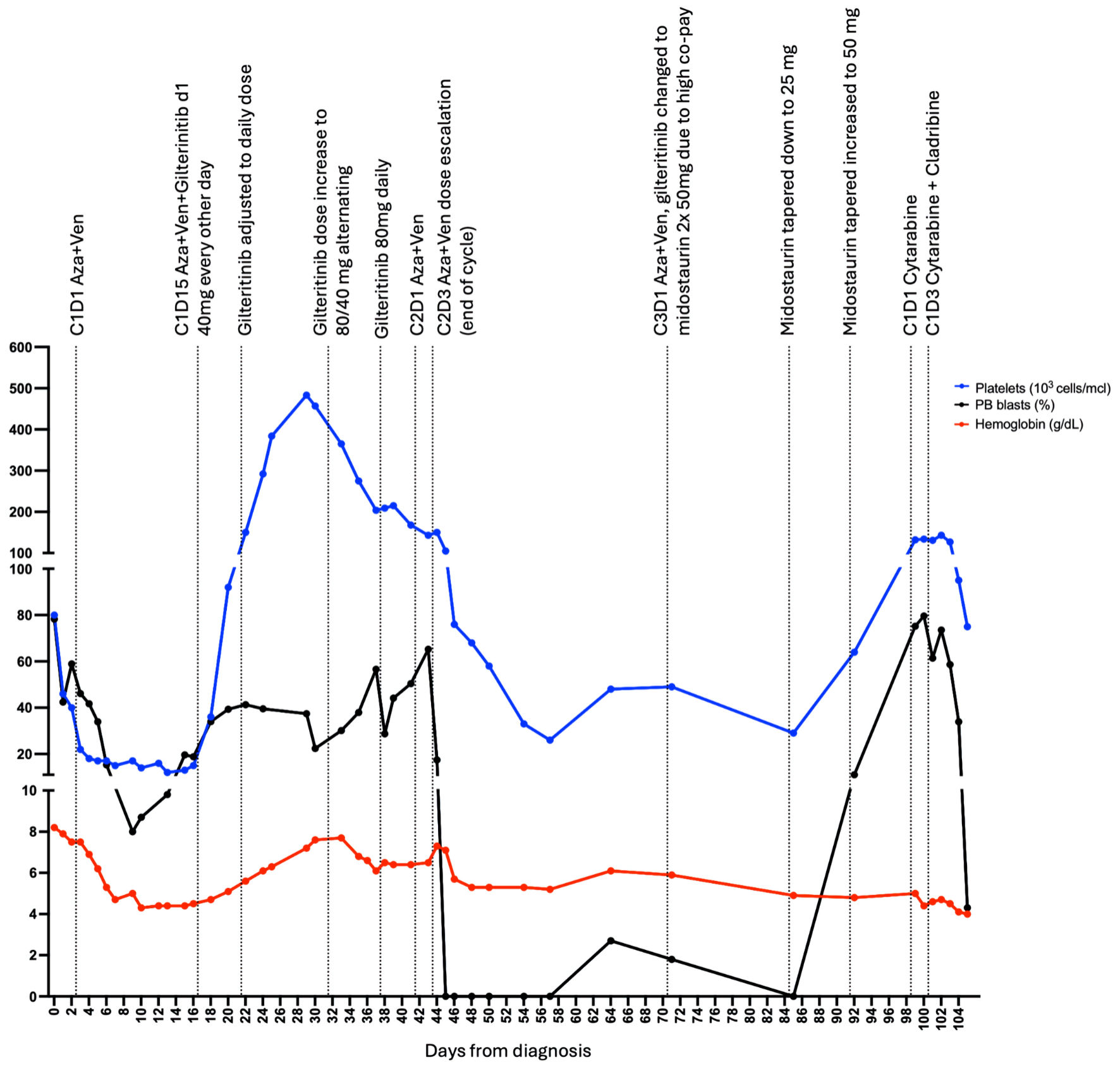
↓ Figure 1. Disease treatment and progression.
Each point represents measurement of peripheral blood (PB) blast percentage (black), hemoglobin level
(red) or platelets measurement (blue). First point represents diagnosis at day 0, C1D1 Aza + Ven
represents cycle one day 1 of the treatment with azacitidine (Aza) and venetoclax (Ven), C1D1 cytarabine
indicates cycle one day 1 of cytarabine treatment, C1D3 cytarabine + cladribine indicates cycle one day
3 of cytarabine and cladribine treatment. Last point represents the last day at the hospital, soon after
the patient expired at a different institution.
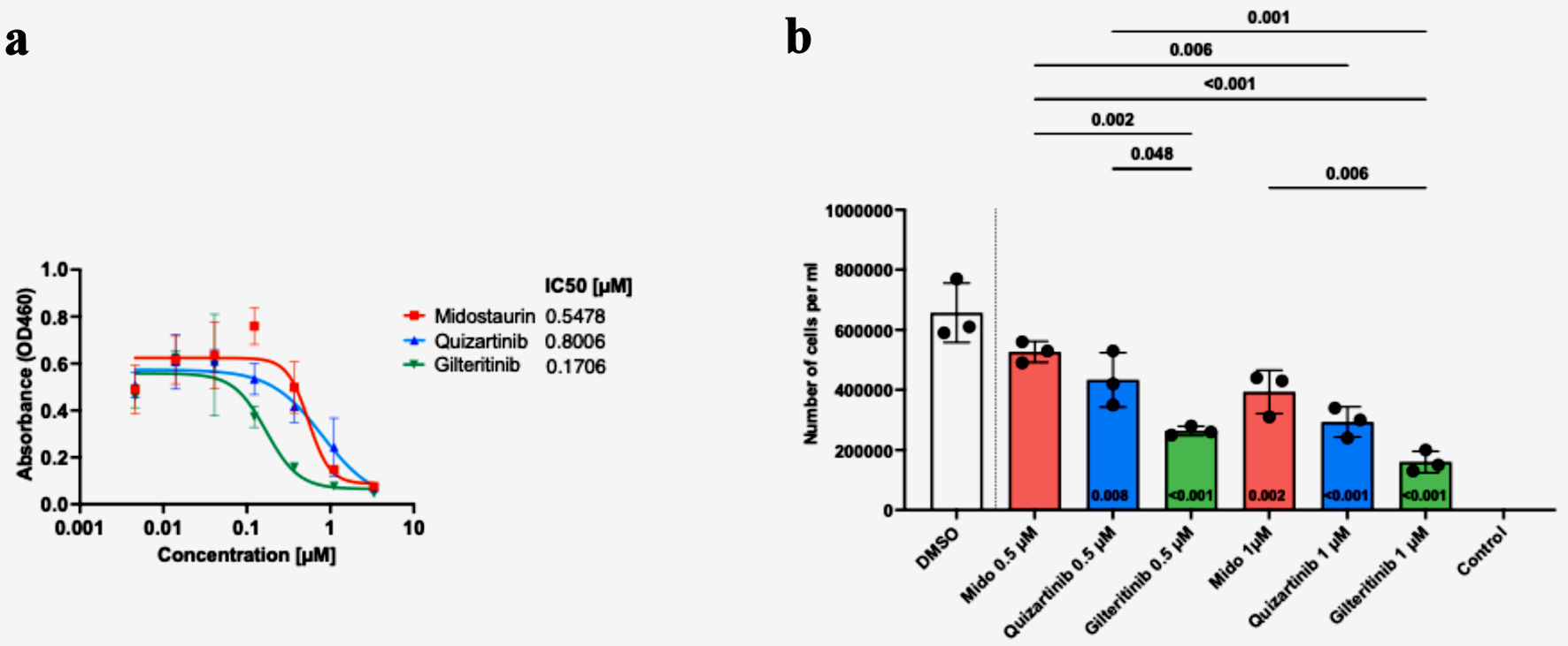
↓ Figure 2. Patient blasts show variable
responses to FLT3 inhibitor treatment, with the highest efficacy of gilteritinib. Cells were seeded with
graded concentrations (0 - 3.33 µM) of midostaurin, quizartinib and gilteritinib for 72 h, and cell
viability was determined by CCK-8 assay. The graph depicts the IC50 values for each drug (a).
Cells were seeded with 0, 0.5, or 1 µM of FLT3 inhibitor or dimethyl sulfoxide (DMSO). Cells were
counted three times 72 h post-treatment with trypan blue (b). Statistical analysis was performed using
one-way ANOVA, with adjusted P < 0.05 considered significant.
↓ Figure 3. Gilteritinib treatment results in
the lowest percentage of live cells among tested FLT3 inhibitors. Flow cytometry analysis of cells
stained with propidium iodide (PI) was performed to evaluate cell death. Cells were first gated for the
lymphocyte population based on side scatter and forward scatter, followed by gating cells on PI-positive
and PI-negative (live cells) populations in reference to unstained control. FLT3: FMS-like tyrosine
kinase 3; DMSO: dimethyl sulfoxide; FSC-A: forward scatter area.
