Journal of Medical Cases, ISSN 1923-4155 print, 1923-4163 online, Open Access
Article copyright, the authors; Journal compilation copyright, J Med Cases and Elmer Press Inc
Journal website https://jmc.elmerpub.com

Case Report

Volume 16, Number 12, December 2025, pages 493-498


Multi-Modality Imaging for Accurate Valvular Lesion Diagnosis: A Case Report of Catastrophic Outcomes From Unrecognized Severe Aortic Regurgitation

Figures

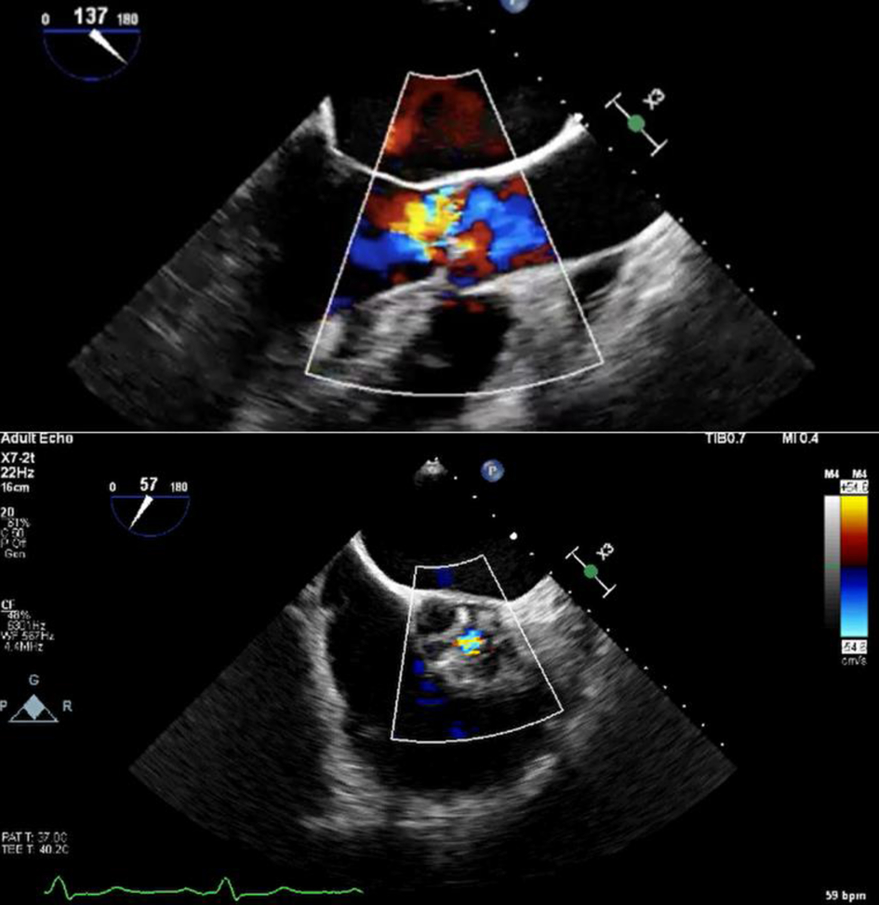
↓  Figure 1. Color Doppler transoesophageal echocardiography showing eccentric aortic regurgitation jet, with short-axis view showing regurgitant jet through coaptation defect.
Figure 1.
↓  Figure 2. Cardiac magnetic resonance imaging with biplane left ventricular volumes and function.
Figure 2.
↓  Figure 3. Cardiac magnetic resonance imaging showing severe aortic regurgitation. Long-axis view showing large central aortic regurgitation jet (white arrow) with dilated left ventricular diameter. Short-axis view showing coaptation failure between the left and right coronary cusps (yellow arrow), contributing to severe aortic regurgitation.
Figure 3.
↓  Figure 4. Cardiac magnetic resonance imaging showing flow across aortic valve. Malalignment of the imaging plane resulted in underestimation of the regurgitant fraction.
Figure 4.