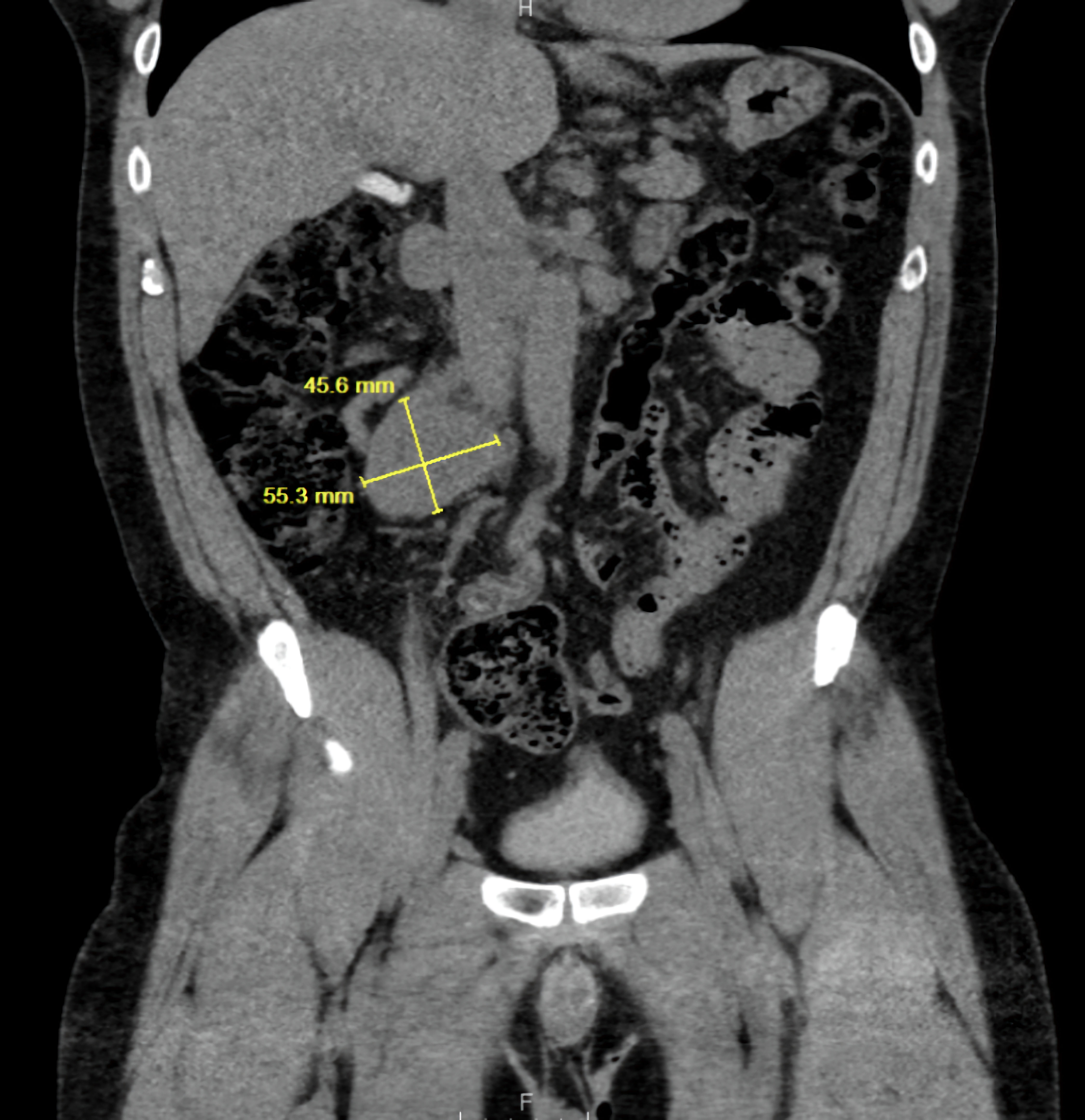
↓ Figure 1. Coronal CT abdomen showing a
retroperitoneal mass measuring 55.3 × 45.0 mm (yellow measurement line), consistent with an
extra-adrenal paraganglioma. CT: computed tomography.
| Journal of Medical Cases, ISSN 1923-4155 print, 1923-4163 online, Open Access |
| Article copyright, the authors; Journal compilation copyright, J Med Cases and Elmer Press Inc |
| Journal website https://jmc.elmerpub.com |
Case Report
Volume 16, Number 8, August 2025, pages 282-286
When the Tumor Leaves but the Damage Lingers: A Case of Delayed Cardiomyopathy Recovery Post-Paraganglioma Resection
Figures