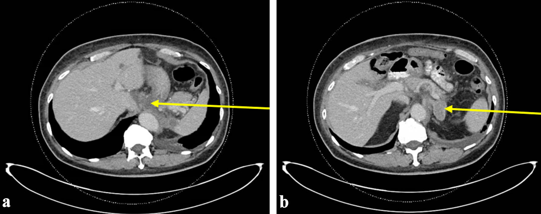
↓ Figure 1. The most recent CT abdomen and pelvis
performed prior to initial ED visit with bleeding. CT scan at this time demonstrated progression of
disease with worsening pulmonary nodules, bilateral retroperitoneal adenopathy, soft tissue thickening
around esophageal hiatus (arrow) (a), and new left adrenal lesion (arrow) (b). ED: emergency department;
CT: computed tomography.
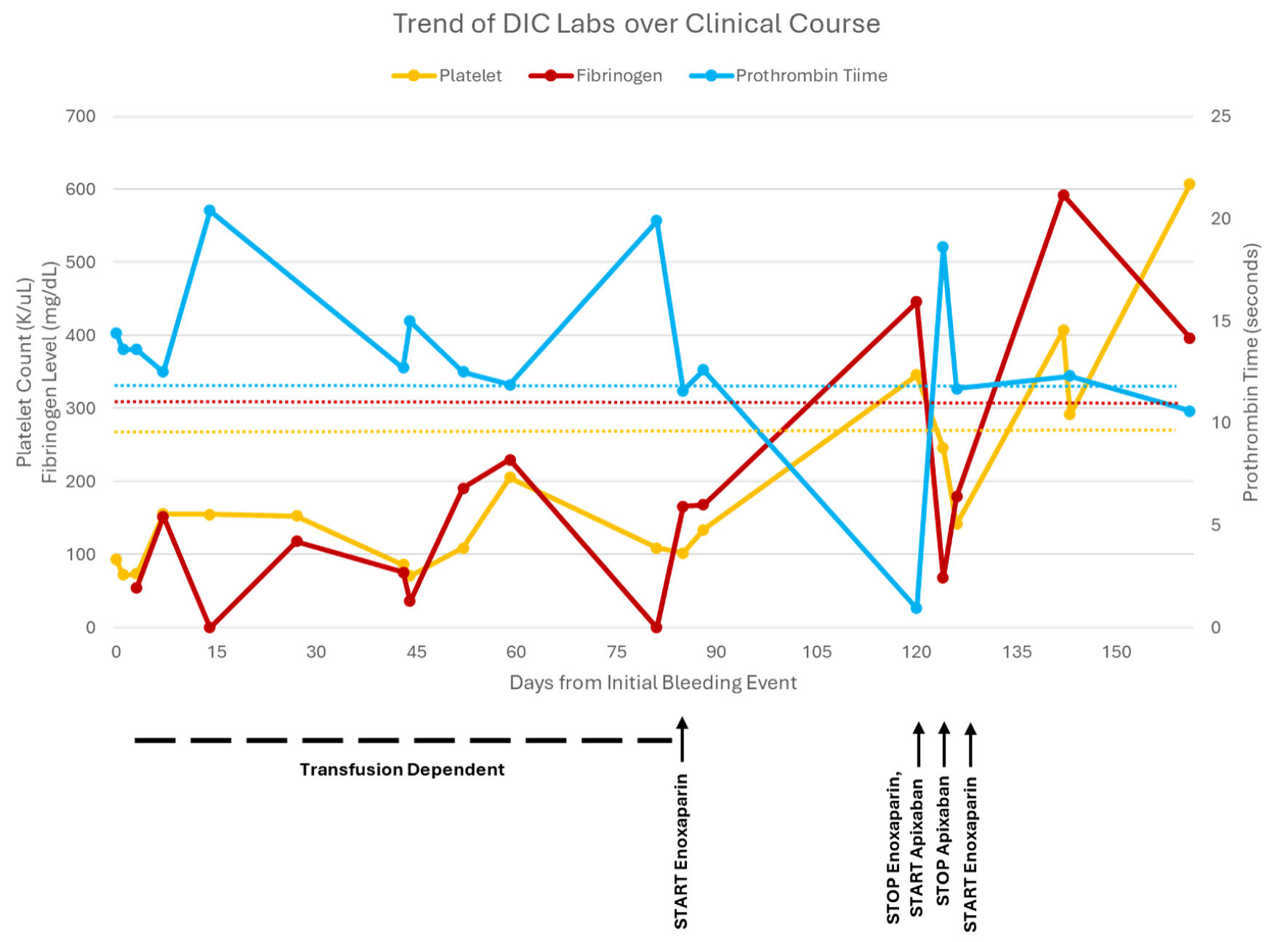
↓ Figure 2. The trend of disseminated
intravascular coagulation (DIC) labs from initial bleeding event until patient death, including platelet
count, fibrinogen level, prothrombin time (PT), and partial thromboplastin time (PTT). PTT was not
included due to simplicity. Additionally, PTT generally correlated with PT except when on enoxaparin and
was higher due to enoxaparin once initiated.
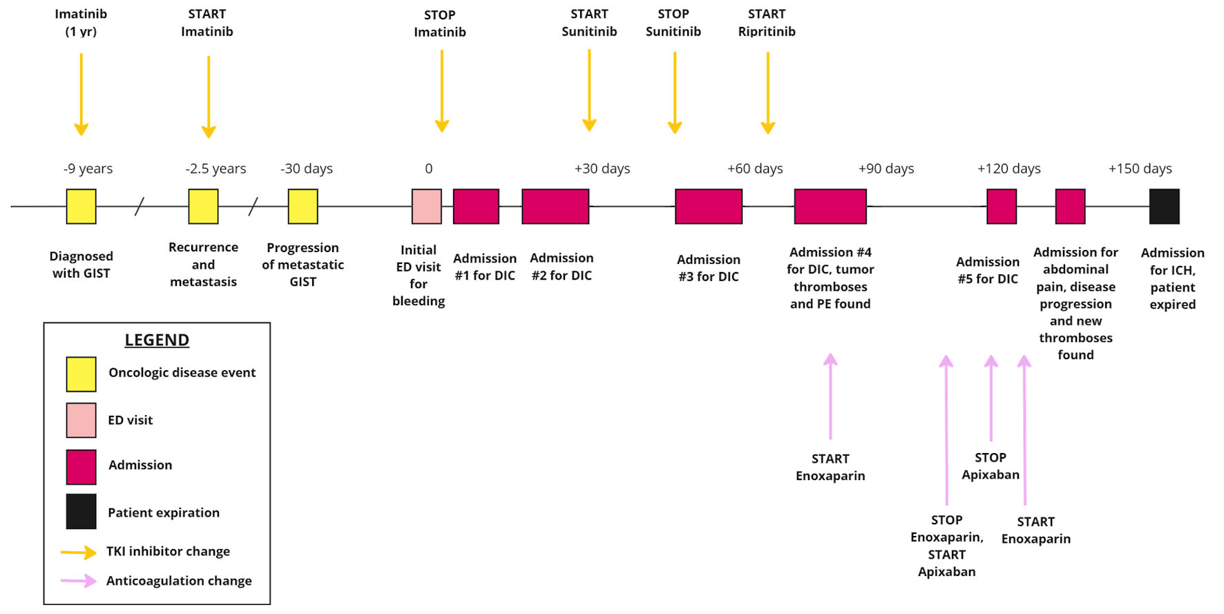
↓ Figure 3. The timeline of patient’s
oncologic disease and progression with subsequent presentations to the hospital, highlighting the
initiation and cessation of TKI inhibitors as well as anticoagulant therapies. GIST: gastrointestinal
stromal tumor; DIC: disseminated intravascular coagulation; ED: emergency department; TKI: tyrosine
kinase inhibitor.
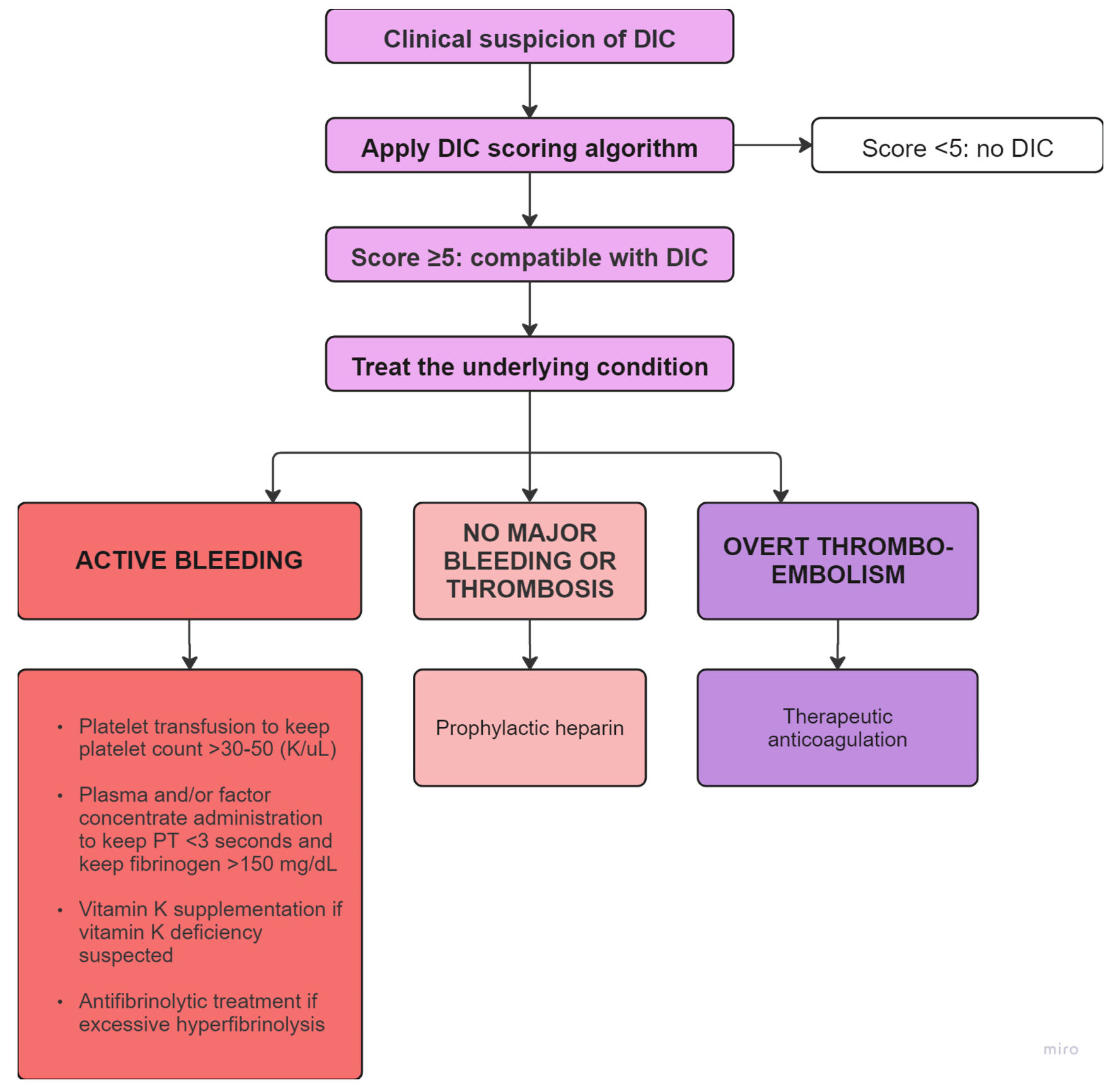
↓ Figure 4. Flowchart depicting the diagnosis and
management of DIC based on bleeding or thromboembolic predominance [1, 18]. This flowchart was adapted
from flowchart developed by Levi et al [1]. PT: prothrombin time; DIC: disseminated intravascular
coagulation.