Journal of Medical Cases, ISSN 1923-4155 print, 1923-4163 online, Open Access
Article copyright, the authors; Journal compilation copyright, J Med Cases and Elmer Press Inc
Journal website https://jmc.elmerpub.com

Case Report

Volume 16, Number 9, September 2025, pages 372-380


Acute Hepatitis in a Patient Treated With Ribociclib for Metastatic Breast Carcinoma

Figures

Figure 1.
Figure 1. CT of the abdomen and pelvis. (a) Axial view demonstrating prominent osteolytic neoplastic lesions within the pelvis (arrows). (b) Coronal view demonstrating prominent osteolytic neoplastic lesions within the spine (arrows). CT: computed tomography.
Figure 2.
Figure 2. Invasive lobular breast carcinoma. (a) Right iliac bone lesion showing metastatic carcinoma (asterisk) (H&E stain, original magnification, × 100). (b) Strong positive immunostaining for CK7 in the metastatic neoplastic cells (arrow) (immunohistochemistry, original magnification, × 200). (c) Focal positive immunostaining for GATA3 in the metastatic neoplastic cells (arrow) (immunohistochemistry, original magnification, × 200). (d) Focal positive immunostaining for mammaglobin in the metastatic neoplastic cells (arrow) (immunohistochemistry, original magnification, × 200). (e) Strong positive immunostaining for GCDFP-15 in the metastatic neoplastic cells (arrow) (immunohistochemistry, original magnification, × 200). (f) Breast mass biopsy showing invasive lobular carcinoma, grade 2 (arrow) (H&E stain, original magnification, × 400). H&E: hematoxylin and eosin.
Figure 3.
Figure 3. Course of liver enzyme and bilirubin elevation and declination in relation to ribociclib administration, discontinuation, and NAC treatment. NAC: N-acetylcysteine.
Figure 4.
Figure 4. Liver biopsy histology. (a) Centrilobular confluent hepatocyte necrosis (asterisk) with mild lymphocytic portal and lobular inflammation (H&E stain, original magnification, × 100). (b) Higher magnification of extensive centrilobular confluent hepatocyte necrosis (arrow) (H&E stain, original magnification, × 200). (c) Individual necrosis (arrows highlighting apoptotic hepatocytes) (H&E stain, original magnification, × 400). (d) Trichrome special stain demonstrating collapsed reticulin fibers (arrows) due to hepatocyte necrosis, it is negative for fibrosis (special stain, original magnification, × 400). (e) Reticulin special stain demonstrating significant hepatic parenchyma collapse (special stain, original magnification, × 400). H&E: hematoxylin and eosin.

Table

Table 1. Causality Assessment Tools Applied to Ribociclib-Induced Liver Injury
 
Assessment toolPurposeInput criteriaPatient-specific resultsInterpretationCase relevance
ADR: adverse drug reaction; ALT: alanine aminotransferase; AST: aspartate aminotransferase; DILI: drug-induced liver injury; RUCAM: Roussel Uclaf Causality Assessment Method; ULN: upper limit of normal.
R-factorClassify type of liver injury (hepatocellular vs. cholestatic vs. mixed)
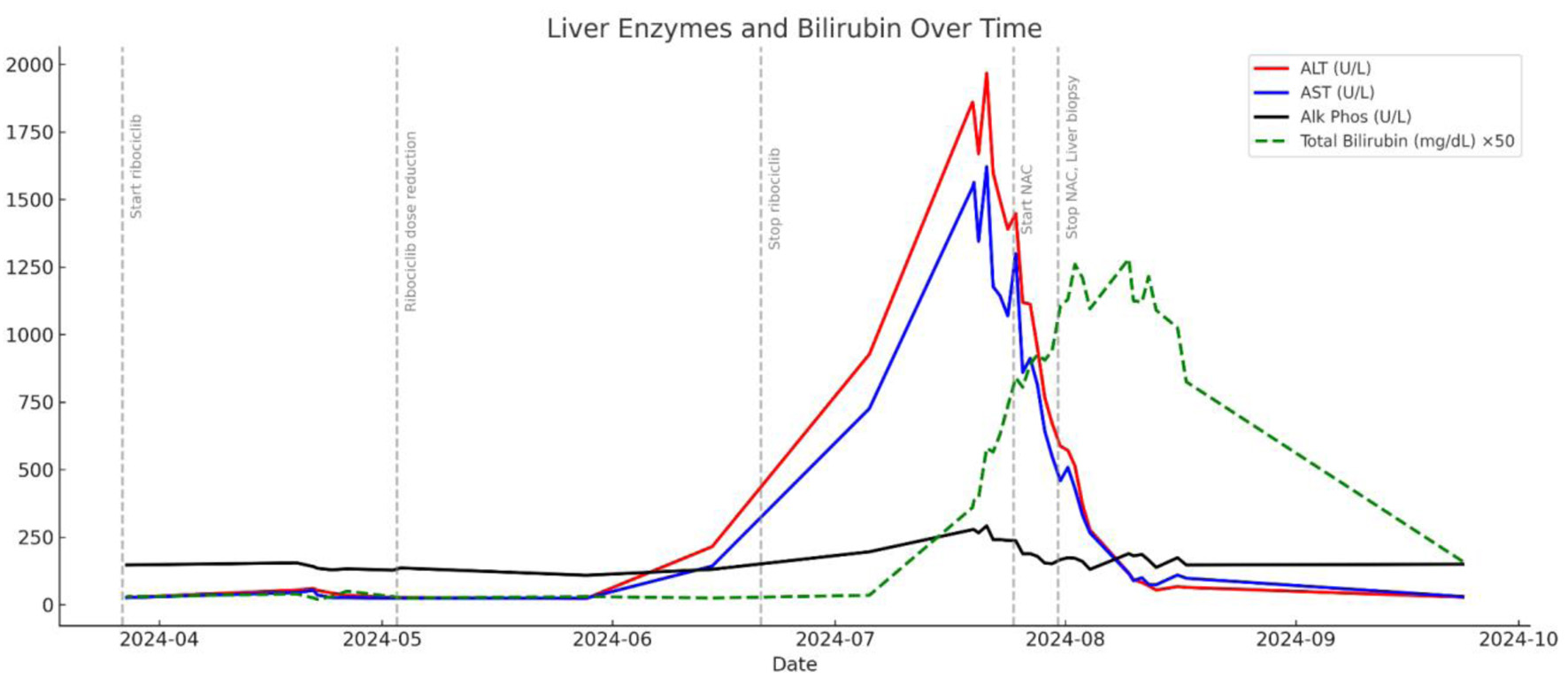
R = (ALT ÷ ALT ULN) ÷ (ALP ÷ ALP ULN); ALT = 1,825 U/L (ULN = 40), ALP = 278 U/L (ULN = 120)R = (1,825/40) ÷ (278/120) = 20.73Hepatocellular injury (R ≥ 5)Confirms hepatocellular pattern, consistent with biopsy findings
RUCAM scoreEstimate likelihood of drug causality in DILITemporal relationship, risk factors, exclusion of alternative causes, dechallenge, known hepatotoxicity, rechallengeComponents (approx): +2 (onset); +3 (dechallenge); +2 (no alt causes); +2 (known hepatotoxin); +1 (female); Total: ∼ 10Highly probable (≥ 9)Strongly supports ribociclib as cause of liver injury
Naranjo algorithmGeneral tool to estimate probability of ADRIncludes timing, prior reports, dechallenge, rechallenge, alternative causesComponents (approx): +2 (timing); +1 (dechallenge); +1 (prior reports); +2 (no alt cause); +1 (biopsy); Total: ∼ 7Probable ADR (5 - 8)Supports ribociclib as likely cause of DILI