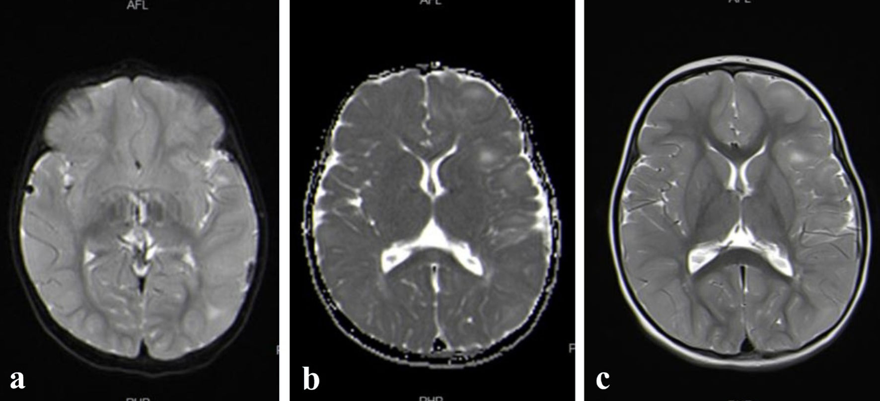
↓ Figure 1. (a, b, c) Brain MRI reported multiple
subependymal nodules, with a mild subtle increase in the enhancement of the lesion on the left centrum
semiovale. MRI: magnetic resonance imaging.
| Journal of Medical Cases, ISSN 1923-4155 print, 1923-4163 online, Open Access |
| Article copyright, the authors; Journal compilation copyright, J Med Cases and Elmer Press Inc |
| Journal website https://jmc.elmerpub.com |
Case Report
Volume 16, Number 9, September 2025, pages 345-351
Renal Cell Carcinoma in A Girl With Tuberous Sclerosis Due to a New Mutation
Figures
Tables
| Minor criteria | Major criteria | Genetic diagnosis |
|---|---|---|
| 1. “Confetti” skin lesions | 1. Hypomelanotic macules (≥ 3, ≥ 5 mm in diameter) | The definite loss of function mutation in TAC1 and/or TSC2 genes |
| 2. Dental enamel pits (> 3) | 2. Facial angiofibromas (≥ 3) or frontal fibrous plaque | |
| 3. Intraoral fibroma (≥ 2) | 3. Ungual fibromas (≥ 2) | |
| 4. Retinal achromic patch | 4. Shagreen patch or multiple collagenoma | |
| 5. Multiple renal cysts | 5. Multiple retinal hamartomas | |
| 6. Non-renal hamartomas | 6. Cortical dysplasia | |
| 7. Subependymal nodules | ||
| 8. Subependymal giant cell astrocytoma | ||
| 9. Cardiac rhabdomyoma | ||
| 10. Pulmonary lymphangioleiomyomatosis | ||
| 11. Renal angiomyolipomas (≥ 2) |
| Study | Age at presentation | Presenting features |
|---|---|---|
| [32] | 17 months | Seizure |
| [33] | 6 months | Seizure |
| [33] | 3 months | Seizure |
| [23] | 3 months | Seizure, delays in speech, cognitive abilities, and social development |
| [23] | 4 months | Seizure |
| [34] | 32 years | Unilateral angiofibroma and hypopigmented patch |
| [35] | 9 years | Unilateral facial angiofibroma |
| [36] | 1 month | Erythema and nodules in the face, neck, and oral cavity, several grain-sized hypopigmentation spots on her back |
| [37] | 7 years | Facial angiofibroma, hypomelanotic macule on trunk, shagreen patch, and tonic clonic convulsion |
| [37] | 3 years | Facial angiofibroma, poliosis (hypomelanosis of hair), and hypomelanotic patches |
| [37] | 7 years | Hypomelanotic patches in face, trunk and limbs |
| [37] | 11 months | Hypomelanotic patch in trunk multiple and focal seizure |