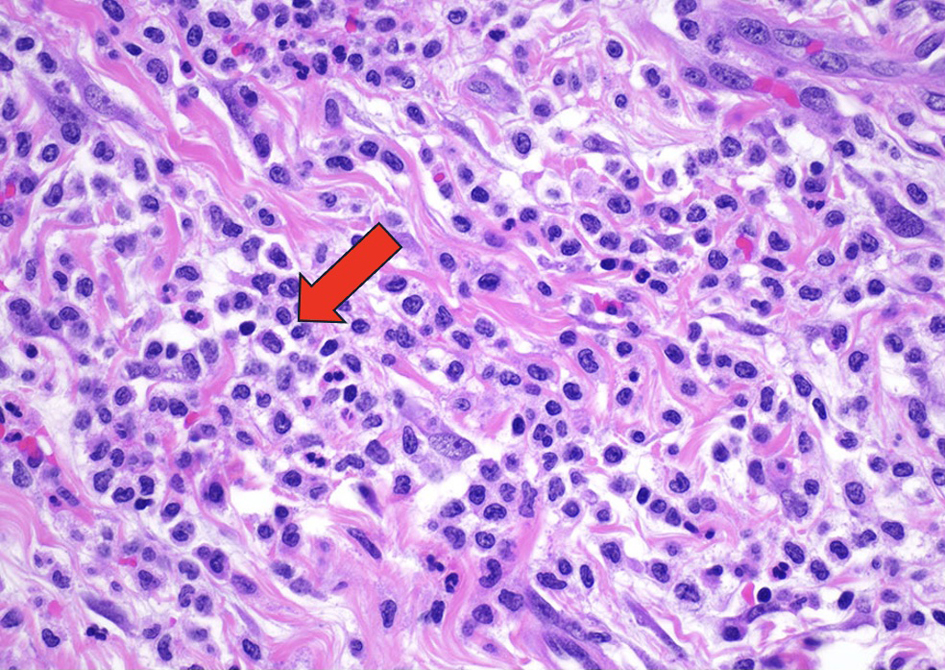
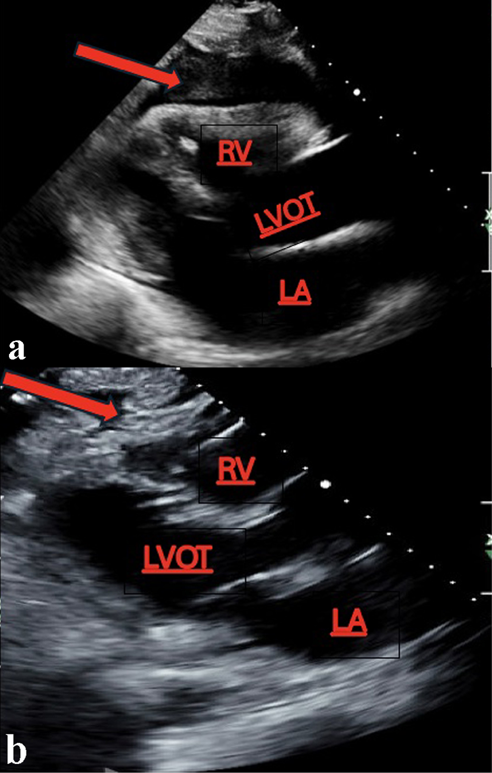
Journal of Medical Cases, ISSN 1923-4155 print, 1923-4163 online, Open Access
Article copyright, the authors; Journal compilation copyright, J Med Cases and Elmer Press Inc
Journal website https://jmc.elmerpub.com

Case Report

Volume 000, Number 000, July 2025, pages 000-000


A Heart of Myeloid: Extramedullary Chronic Myelomonocytic Leukemia-2 Presenting as a Myeloid Sarcoma of the Pericardium Causing Recurrent Pericardial Effusions

Figures

Figure 1.
Figure 1. The myocardium and surrounding 
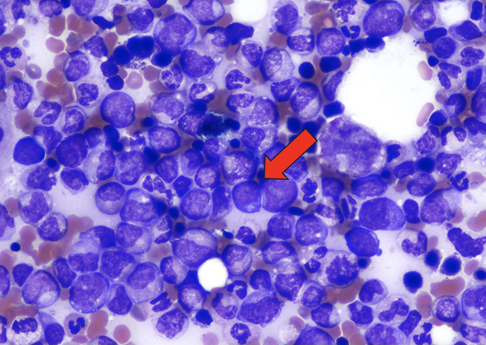
adipose tissue are architecturally effaced by an infiltrating lesion composed of immature mononuclear cells demonstrating fine chromatin. Hematoxylin and eosin (H&E)-stained sections of the pericardium demonstrated fibrovascular and fibroadipose tissue with a variably dense infiltration of medium sized cells with irregular nuclear membranes, conspicuous nucleoli, and variable amounts of clear cytoplasm, consistent with atypical/immature monocytic cells. These atypical cells formed diffuse sheets in some areas and were seen with large numbers of mature granulocytes in other areas. The immature cells 
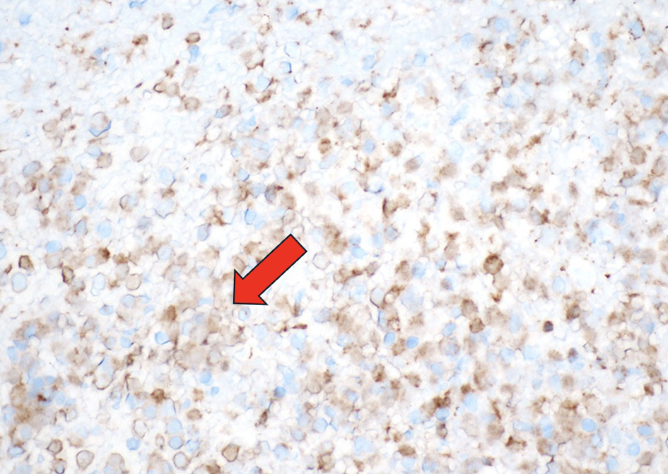
demonstrated positive staining for CD45, CD43, CD34, CD68, and CD163. They were negative for CD117, TdT, CD3, CD20, CD56, calretinin, and WT1. The Ki-67 proliferation index was variably elevated. This atypical monocytic infiltration is diagnostic of myeloid sarcoma (H&E, × 400). Arrow indicating immature mononuclear cells infiltrating the cardiac tissue.
Figure 2.
Figure 2. The immature mononuclear cells stain positive for myeloperoxidase, confirming the diagnosis of myeloid sarcoma (myeloperoxidase, × 400). Arrow indicating the light gold staining of the cells positive for myeloperoxidase.
Figure 3.
Figure 3. Wright Giemsa stain of the bone marrow demonstrating myeloblasts (× 1,000). Arrow indicating myeloblasts.
Figure 4.
Figure 4. (a) A large pericardial effusion with RV collapse clinically correlating with cardiac tamponade. (b) Resolution of the pericardial effusion status after pericardiocentesis. Arrows indicating the fluid collection around the pericardium with RV collapse (a) with subsequent resolution after drainage (b). LA: left atrium; LVOT: left ventricular outflow tract; RV: right ventricle.