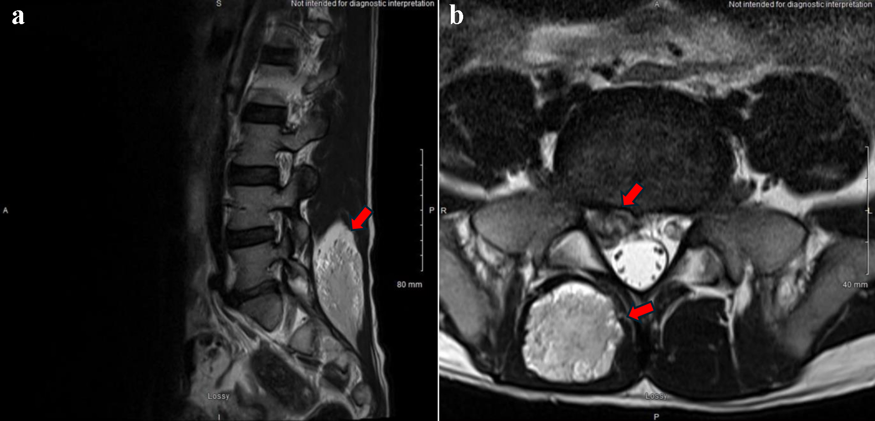
↓ Figure 1. (a) Axial and (b) sagittal
T2-weighted MRI show a well-defined mass at L4/L5 extending to L5/S1 (arrows). MRI: magnetic resonance
imaging.
| Journal of Medical Cases, ISSN 1923-4155 print, 1923-4163 online, Open Access |
| Article copyright, the authors; Journal compilation copyright, J Med Cases and Elmer Press Inc |
| Journal website https://jmc.elmerpub.com |
Case Report
Volume 16, Number 6, June 2025, pages 232-237
Paraspinal Intramuscular Hemangioma at L5-S1 With Concurrent Disc Herniation
Figures
Table
| Source | Age | Sex | Location | Muscle(s) involved | Size | Imaging | Clinical presentation | Outcome |
|---|---|---|---|---|---|---|---|---|
| 18F-FDG-PET: 18F-fluorodeoxyglucose positron emission tomography; MRI: magnetic resonance imaging; US: ultrasound. | ||||||||
| [11] | 29 years | Female | Trunk | Postero-lateral most muscles of right posterior para-spinal muscles | 5.9 × 4.9 × 8.5 cm | US/MRI | A 9 × 7 cm non-tender, soft paraspinal swelling at L3-L4 on the right side became more prominent with painful forward and side bending movements, occurring in the setting of lower back pain and reduced lumbar lordosis but without bony tenderness or neurological findings. | Mass was excised. Symptomless local recurrence was found during a routine follow-up. MRI was done after 2 years. |
| [12] | 39 years | Male | Trunk | Not reported | Not reported | Not reported | Asymptomatic subcutaneous mass which had been present for 3 years | No recurrence was seen for 1 year following excision. |
| [13] | 37 years | Male | Trunk | Paraspinal/iliopsoas | 4.2 × 2 × 3.1 cm | US | Painless, started 9 months prior and was stable in size | Not reported |
| [13] | 19 years | Female | Trunk | Erector spinae | 8 × 5 × 3 cm | MRI/US | Painless, started 2 months prior and was stable in size | Not reported |
| [14] | 36 years | Female | Left buttock | Gluteus medius | 15 × 10 × 5 cm | X-ray/MRI | A 5-year history of incidental and infrequent pain of the left buttock accompanied by radiating pain in the back of the thigh | Eleven months after the operation, the manual muscle testing score of her gluteus medius muscle was 5, and she resumed her normal work and exercise levels with no recurrence of pain. |
| [15] | 20 years | Male | Trunk | Longissimus and multifidus muscle | Approximately 4 cm | MRI and 18F-FDG-PET | Back pain and tenderness on upper lumbar level. Additionally, he had compression fractures of the L1, L2, and L3 vertebral bodies, as well as the spinous process fracture at L2. | Not reported |