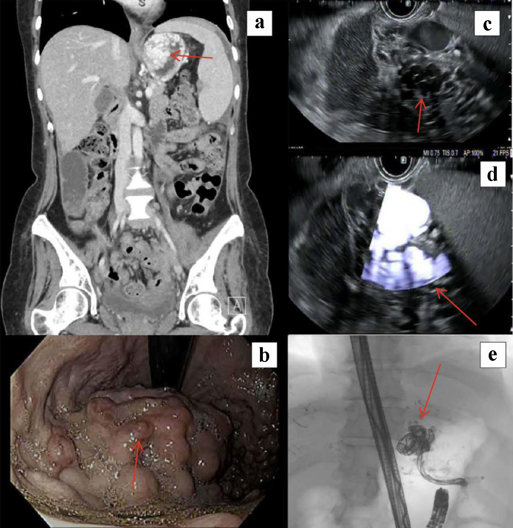
↓ Figure 1. (a) Computed tomography of the
abdomen/pelvis demonstrating splenomegaly and dilated collaterals in the proximal stomach (arrow). (b)
Upper endoscopy with high-risk variceal stigmata (red wale signs, arrow). (c) Endoscopic ultrasound
sonographic image of variceal bundle in gastric wall (arrow). (d) Doppler image of variceal bundle
demonstrating significant inflow (arrow). (e) Fluoroscopic image post-embolization. Visible vascular
coils and lipiodol mixture within the body of the largest variceal bundle as well as the inflow tract
from the splenic hilum (arrow).