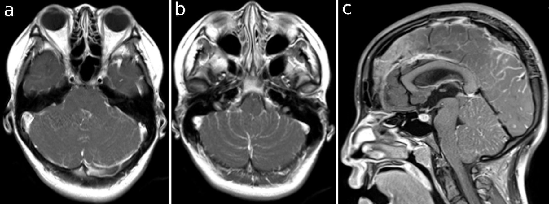
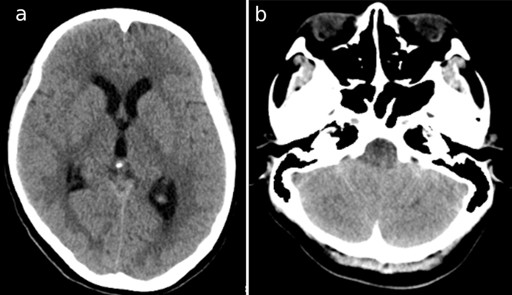
Journal of Medical Cases, ISSN 1923-4155 print, 1923-4163 online, Open Access
Article copyright, the authors; Journal compilation copyright, J Med Cases and Elmer Press Inc
Journal website https://jmc.elmerpub.com

Case Report

Volume 16, Number 4, April 2025, pages 135-139


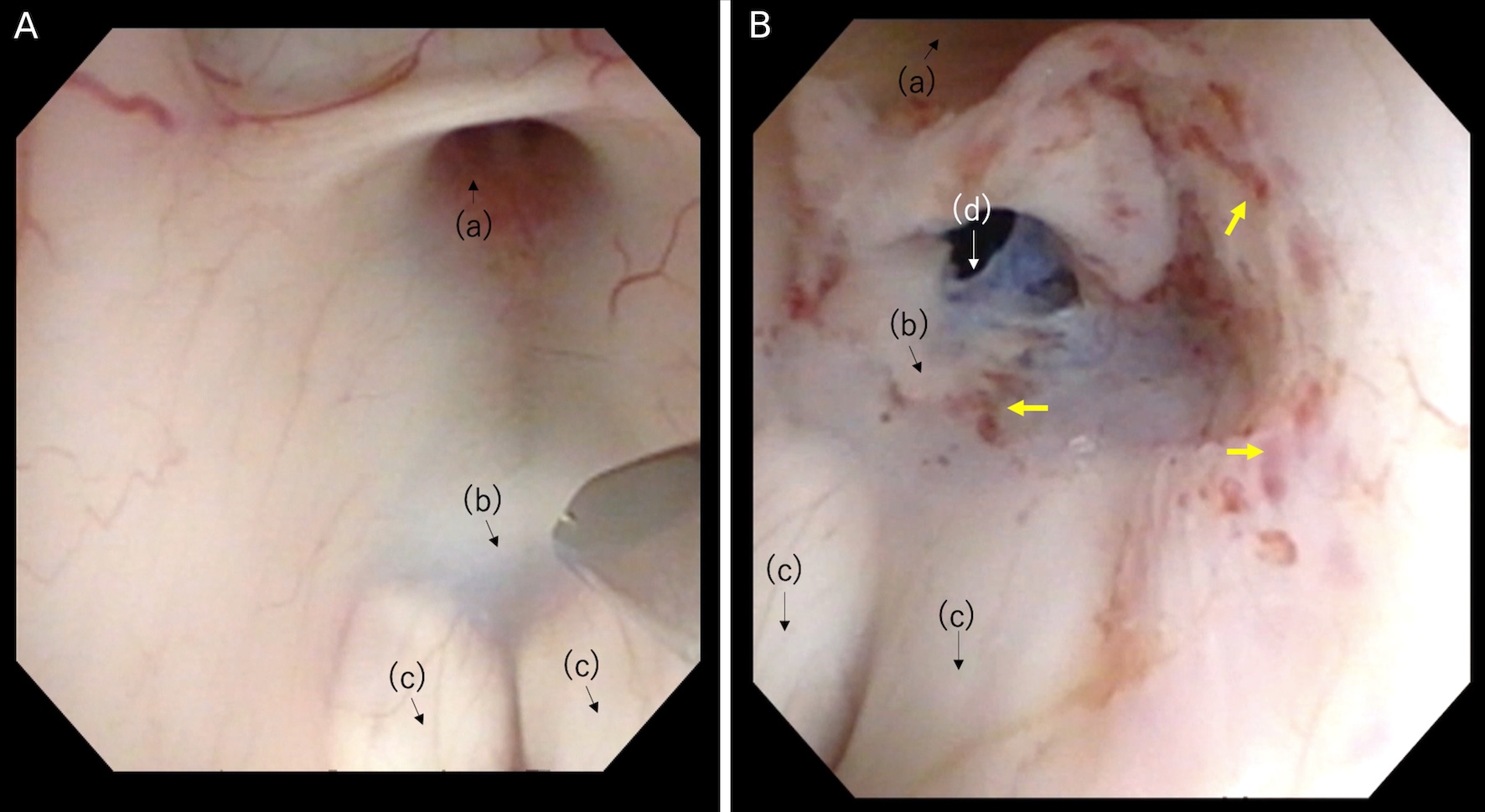
Noncommunicating Hydrocephalus Caused by Aseptic Meningitis Can Be Treated With Endoscopic Third Ventriculostomy

Figures

Figure 1.
Figure 1. CT images at the second admission show (a) the dilatation of bilateral and third ventricles; and (b) cerebellar swelling. CT: computed tomography.
Figure 2.
Figure 2. (a-c) T1 images with gadolinium-enhancing occipital lobe and cerebellar surface, foramina of Magendie and Luschka.
Figure 3.
Figure 3. (A) Thick tuber cinereum before puncture. (B) After the expansion of the stoma on the floor of the third ventricle. Anatomical structures: (a) infundibular recess; (b) tuber cinereum; (c) mamillary bodies; (d) stoma. The yellow arrows show dot bleeding by mechanical injury.
Figure 4.
Figure 4. (a-c) T1 images with gadolinium 1 month after the surgery, demonstrating the improvement of cerebellar edema and shrinkage of ventricles. There was no enhancement of the foramina of Magendie and Luschka. The yellow arrow shows the stoma perforated in ETV.