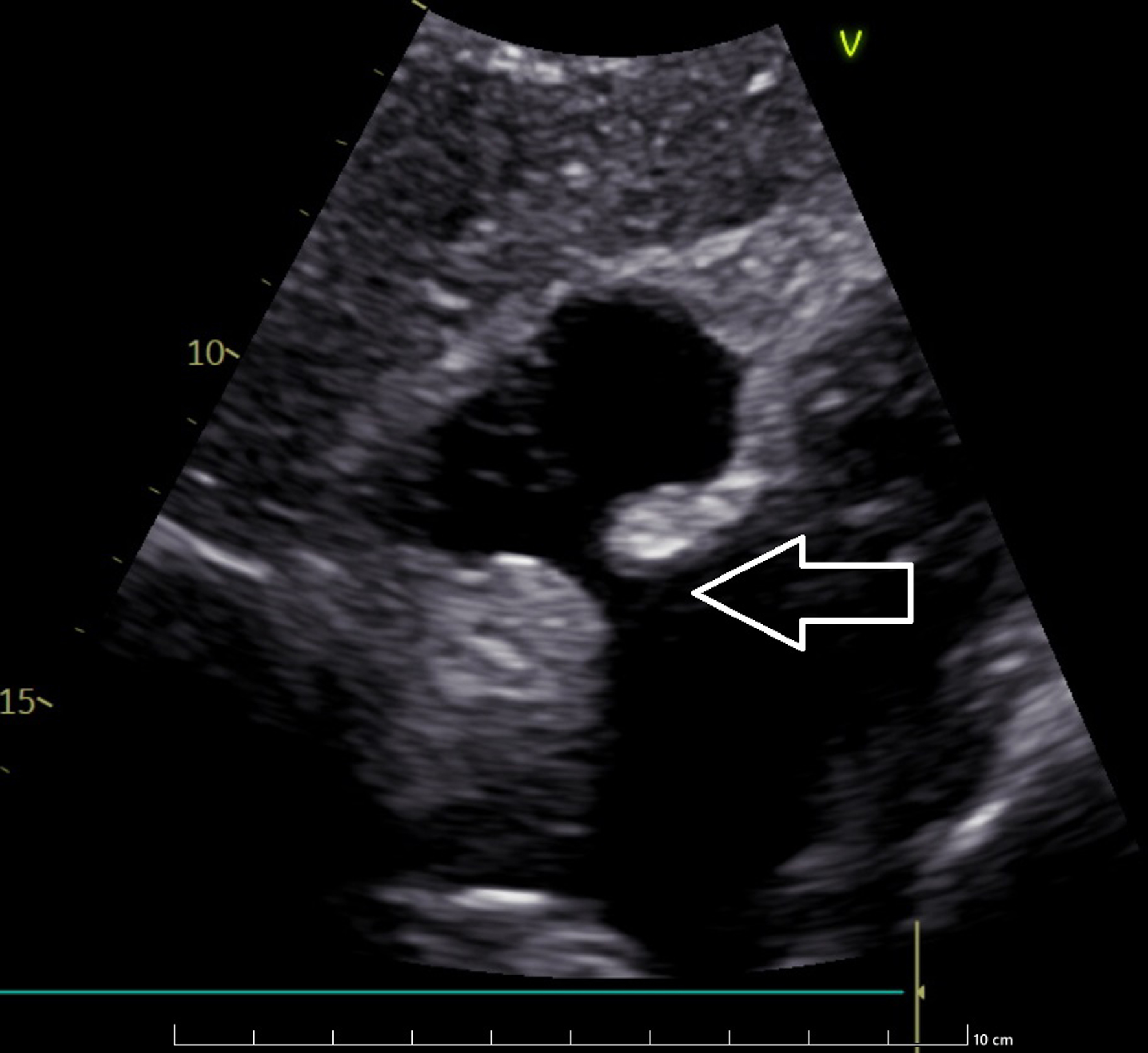
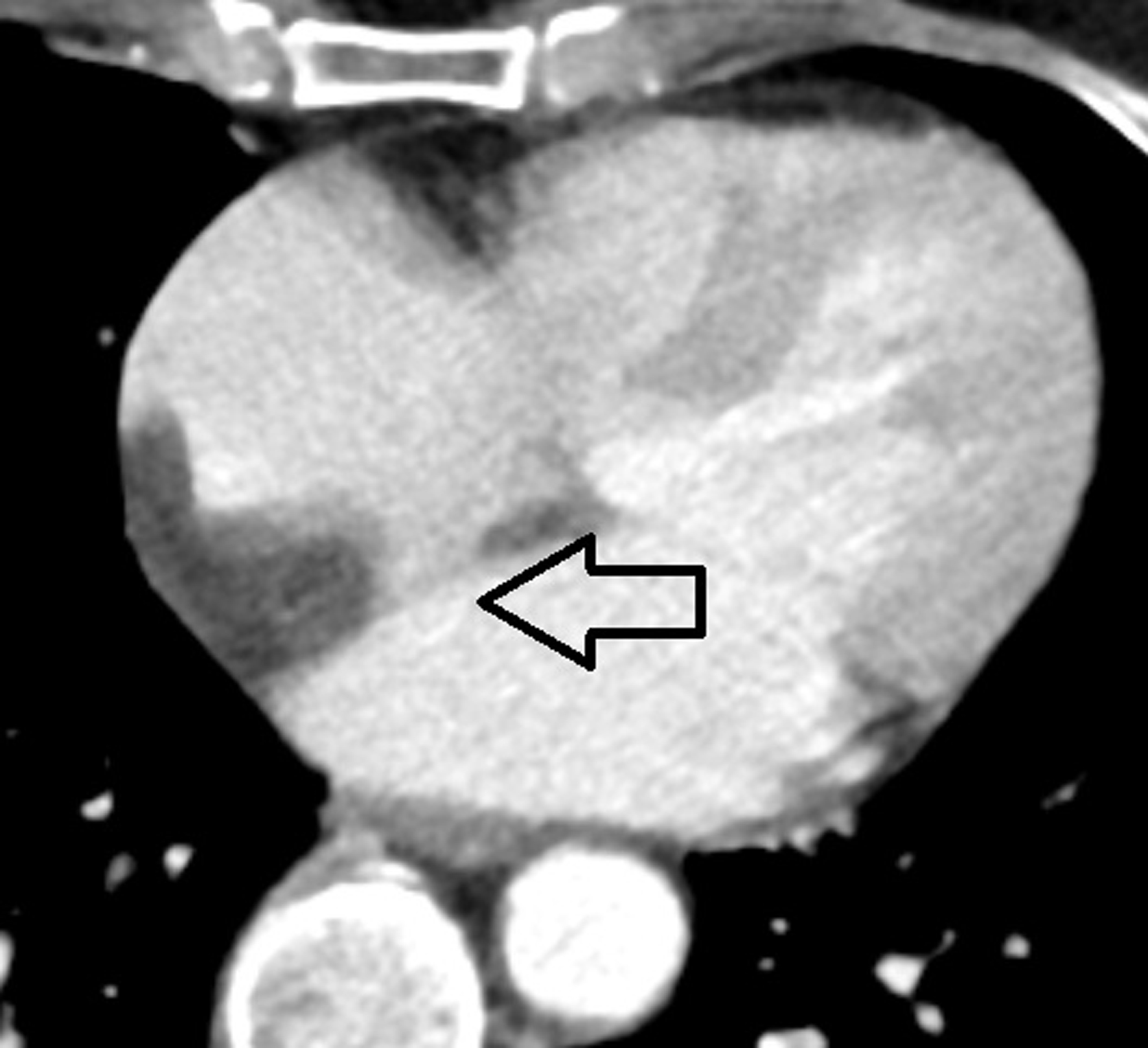
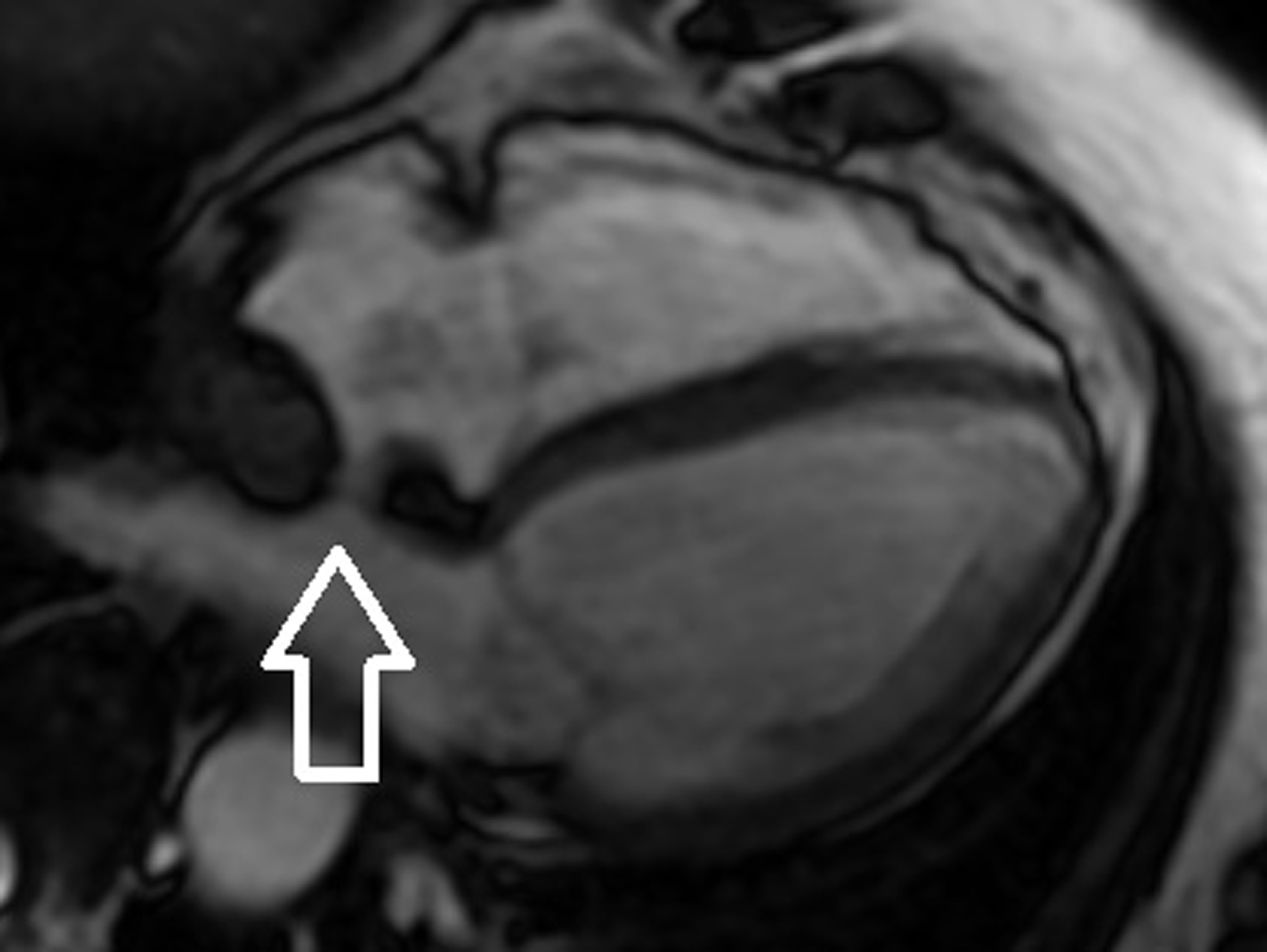
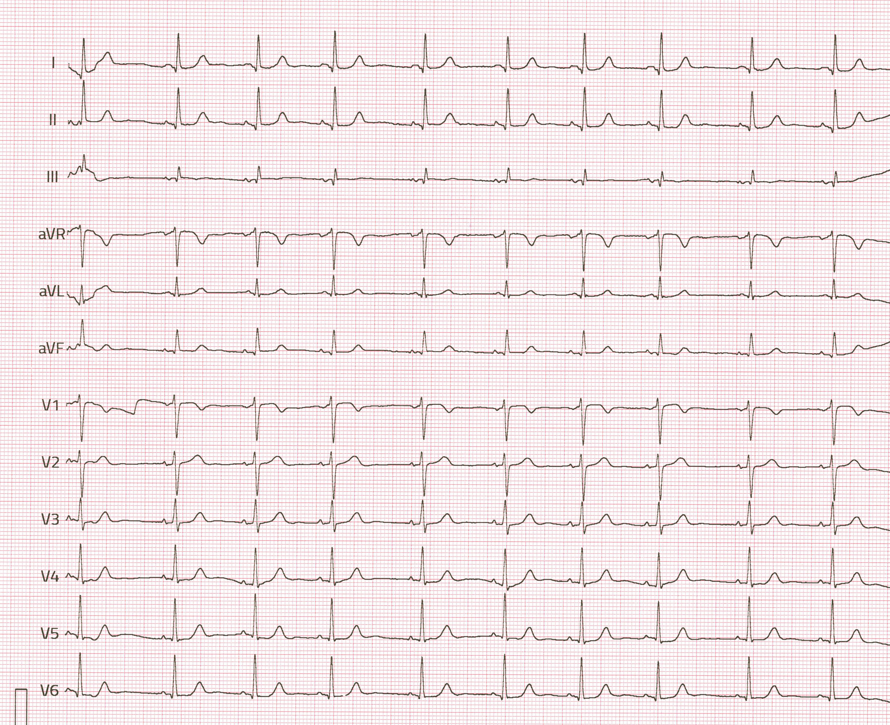
↓ Figure 1. Electrocardiography (ECG) recording
of the patient with lipomatous hypertrophy of interatrial septum (LHIS).
| Journal of Medical Cases, ISSN 1923-4155 print, 1923-4163 online, Open Access |
| Article copyright, the authors; Journal compilation copyright, J Med Cases and Elmer Press Inc |
| Journal website https://jmc.elmerpub.com |
Case Report
Volume 16, Number 3, March 2025, pages 120-126
Lipomatous Hypertrophy of the Interatrial Septum
Figures