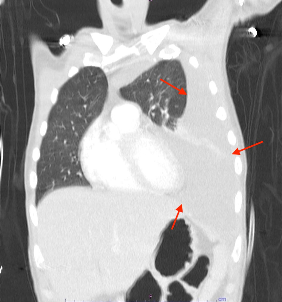
Journal of Medical Cases, ISSN 1923-4155 print, 1923-4163 online, Open Access
Article copyright, the authors; Journal compilation copyright, J Med Cases and Elmer Press Inc
Journal website https://jmc.elmerpub.com

Case Report

Volume 16, Number 2, February 2025, pages 48-54


An Unusual Case of Extracavitary/Solid Variant Primary Effusion Lymphoma With Associated Hemophagocytic Lymphohistiocytosis

Figures

Figure 1.
Figure 1. CT of the chest with contrast demonstrating an 8.7 × 7.1 × 4 soft tissue mass extending along the middle and posterior mediastinum into the left hilum (arrow). The lesion is exerting mass effect on the left atrium, encasing several pulmonary veins, and effacing the fat plane along the anterior esophagus with esophageal wall thickening. CT: computed tomography.
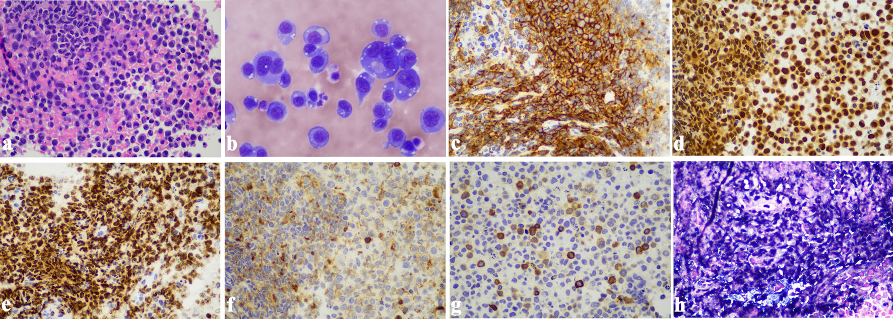
Figure 2.
Figure 2. CT thorax with contrast demonstrating a large pleural effusion occupying the majority of the left hemithorax (arrows). CT: computed tomography.
Figure 3.
Figure 3. Primary effusion lymphoma. (a) Sheets of large pleomorphic lymphoma cells with prominent nucleoli and abundant cytoplasm, morphologically consistent with EC-PEL (H&E stain, original magnification: × 400). (b) Cytospin smear of pleural fluid showing PEL (Wright-Giemsa stain, original magnification: × 600). (c) Strong positive immunostaining for CD138 in neoplastic cells (immunohistochemistry, original magnification: × 400). (d) Strong positive immunostaining for MUM-1 in neoplastic cells (immunohistochemistry, original magnification: × 400). (e) The lymphoma cells demonstrate strong positive immunostaining for HHV8 (immunohistochemistry, original magnification: × 400). (f) Weak positive immunostaining for CD45 in neoplastic cells (immunohistochemistry, original magnification: × 400). (g) A small subset of neoplastic cells showed positive immunostaining for CD3 (immunohistochemistry, original magnification: × 400). (h) In situ hybridization for EBER was positive in the lymphoma cells (original magnification: × 400). EBER: Epstein-Barr virus-encoded RNA; EC-PEL: extracavitary primary effusion lymphoma; H&E: hematoxylin and eosin stain; PEL: primary effusion lymphoma.

Tables

Table 1. Patient HScore
 
CriteriaPresent casePoints
HIV: human immunodeficiency virus; AST: aspartate aminotransferase.
Known underlying immunosuppressionYes (HIV positive)18
Temperature38.9 °C33
OrganomegalyHepatomegaly and splenomegaly38
Number of cytopenias3 lineages34
Ferritin14,134 ng/mL50
Triglycerides298 mg/dL44
Fibrinogen264 mg/dL0
AST16519
HScore (total points)236
Probability of hemophagocytic syndrome based on HScore98-99%


Table 2. HLH-2004 Criteria in Present Case
 
CriteriaPresent case
HLH: hemophagocytic lymphohistiocytosis; IL: interleukin.
FeverYes (38.9 °C)
SplenomegalyYes
Cytopenia (affecting ≥ 2 of 3 lineages in peripheral blood)Yes
Hypertriglyceridemia and/or hypofibrinogenemia (fasting triglycerides > 3.0 mmol/L (> 265 mg/dL) or fibrinogen ≤ 1.5 g/L)Triglycerides: 298 mg/dL
Hemophagocytosis in bone marrow, spleen, liver, lymph nodes, or other tissuesNot identified in bone marrow aspirate
Low or absent natural killer (NK) cell activityNot measured
Serum ferritin concentration ≥ 500 µg/L14,134 µg/L
Soluble CD25 (soluble IL-2 receptor) ≥ 2,400 U/mL39,950 U/mL
Number of criteria fulfilled6 out of 8 (at least 5 criteria needed to fulfill HLH diagnosis)