Figures
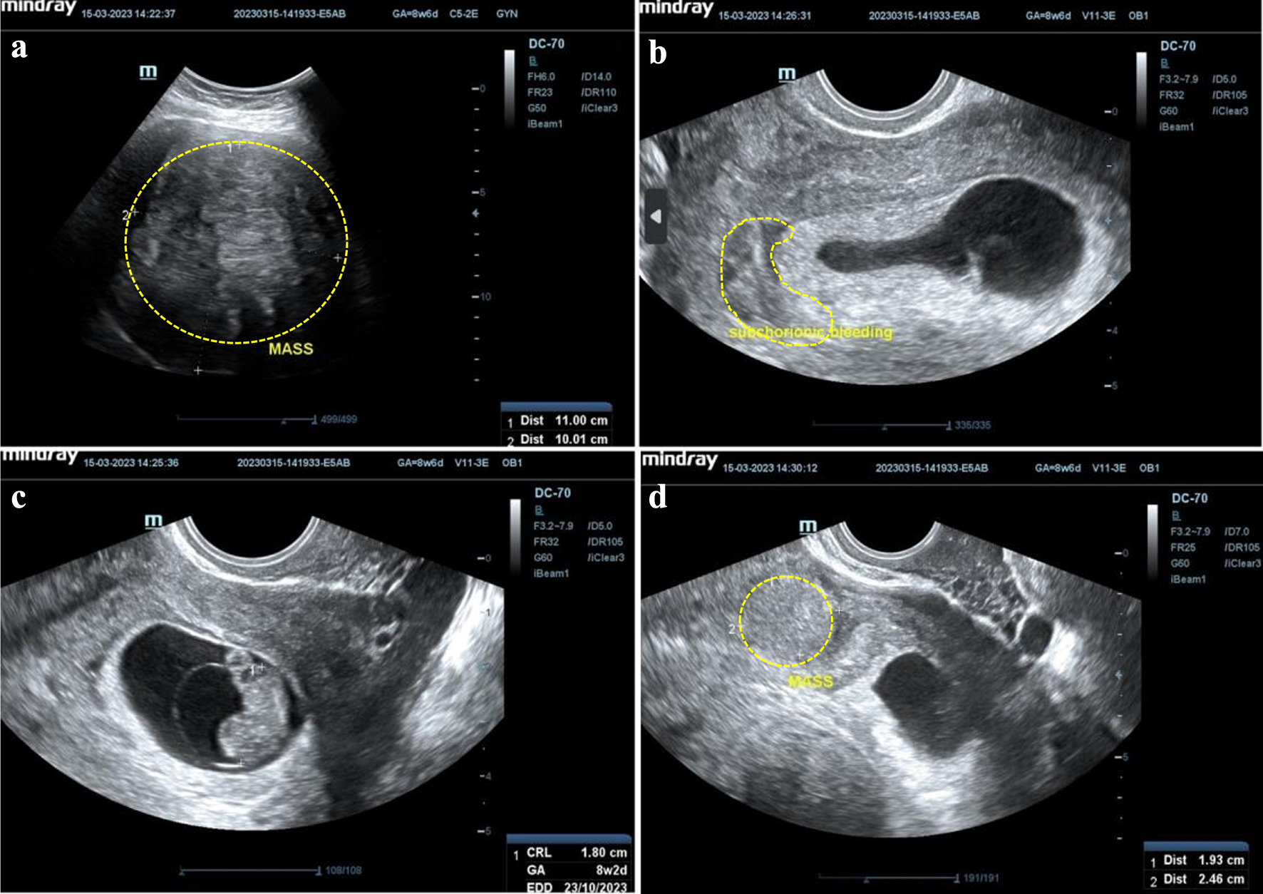
↓ Figure 1. Initial ultrasonography examination.
(a) A mass, at fundal uterine, hyperechoic density, measuring 11 × 10 cm. (b) Subchorionic
bleeding. (c) Single, live fetus, intrauterine, crown-rump length (CRL) 1.8 cm according to 8 weeks and
2 days of pregnancy, positive fetal heart movement. (d) A mass, at isthmus site, hyperechoic density,
measuring 5.46 × 6.27 cm.
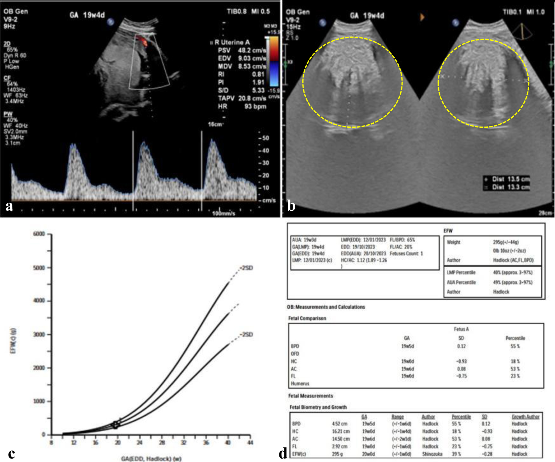
↓ Figure 2. Initial fetomaternal ultrasonography
examination. (a) Right uterine artery pulsatility index (PI), resistance index (RI), and
systolic/diastolic (S/D) ratio of 1.91, 0.81, and 5.34, negative notching. (b) A mass, at posterior
corpus, hyperechoic density, measuring 13 × 13 cm. (c, d) Lubchenco curve, gestational age (GA) and
estimated fetal weight (EFW) show normal growth lines.
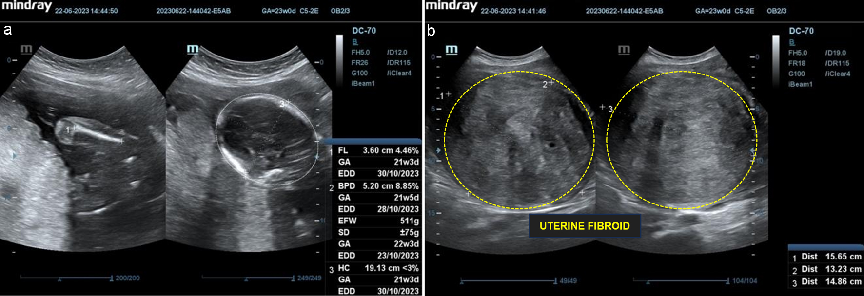
↓ Figure 3. Follow-up ultrasonography examination
on second trimester. (a) Single, live fetus, intrauterine, biometric according to 21 - 22 weeks of
pregnancy, estimated fetal weight (EFW) 511 g, positive fetal heart rate. (b) A mass, at posterior
corpus, hyperechoic density, measuring 15.65 × 13.23 × 14.86 cm, negative
neovascularization.
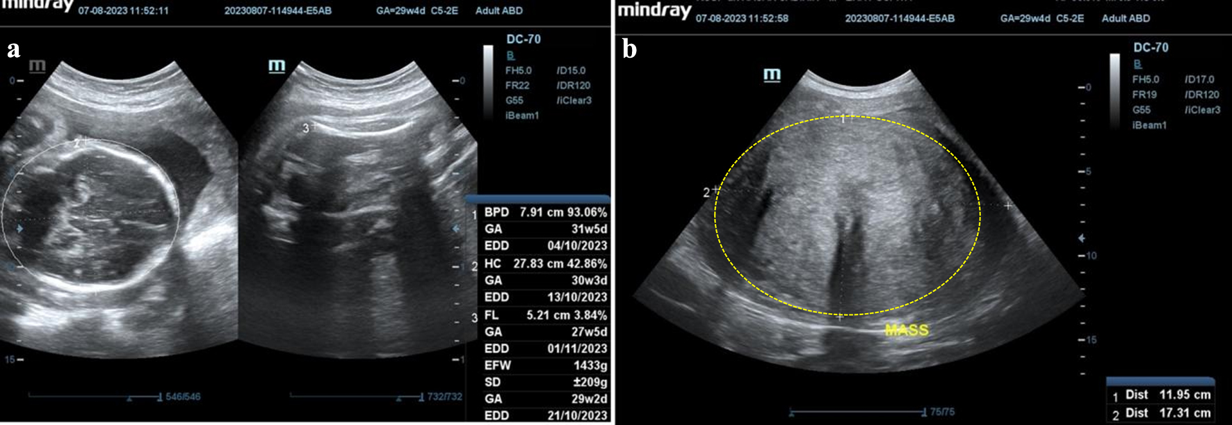
↓ Figure 4. Follow-up ultrasonography examination
on third trimester. (a) Single fetus, live, intrauterine, biometric according to 30 - 31 weeks of
pregnancy, estimated fetal weight (EFW) 1,433 g, positive fetal heart rate. (b) A mass, at posterior
corpus, hyperechoic density, measuring 11.95 × 17.31 × 14.86 cm, negative
neovascularization.
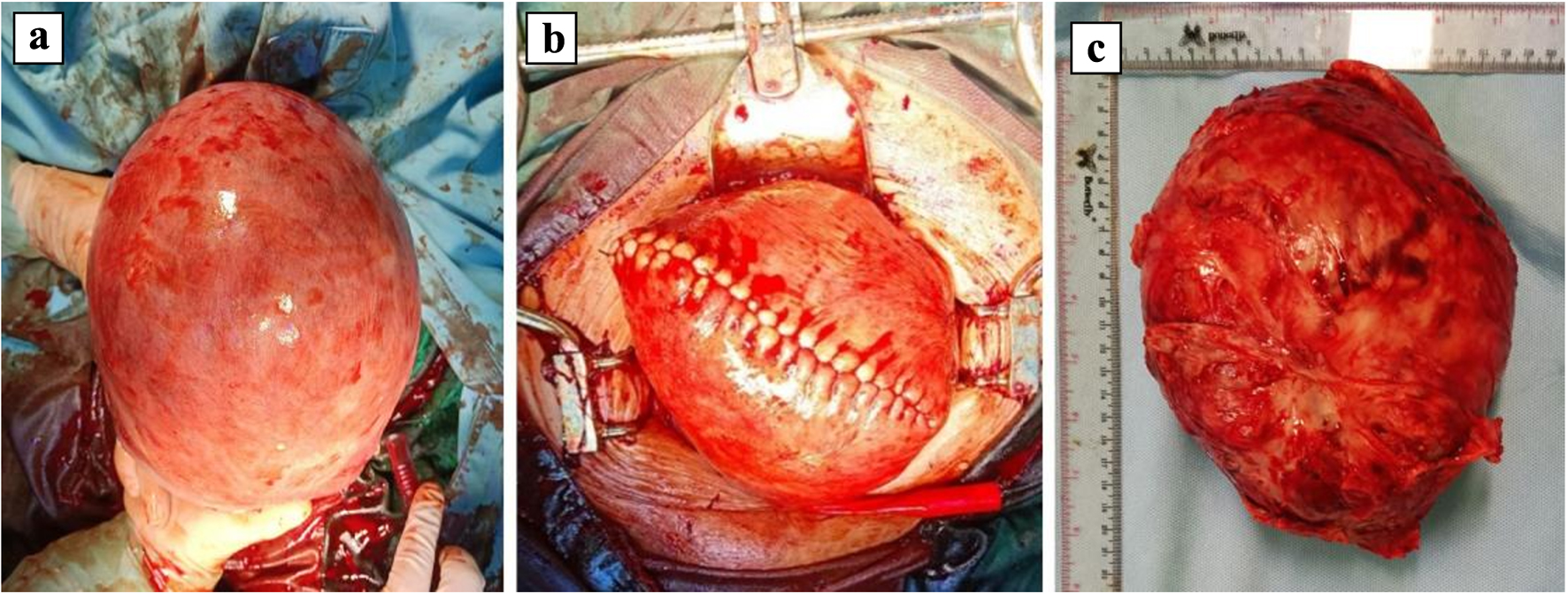
↓ Figure 5. Intraoperative findings showing a
large uterine fibroid mass. (a) The uterine view after delivery, inferior view, a mass at fundal,
measuring around 20 × 15 × 10 cm. (b) The uterine view after enucleation and reparation,
superior view. (c) The specimen mass after enucleation procedure.