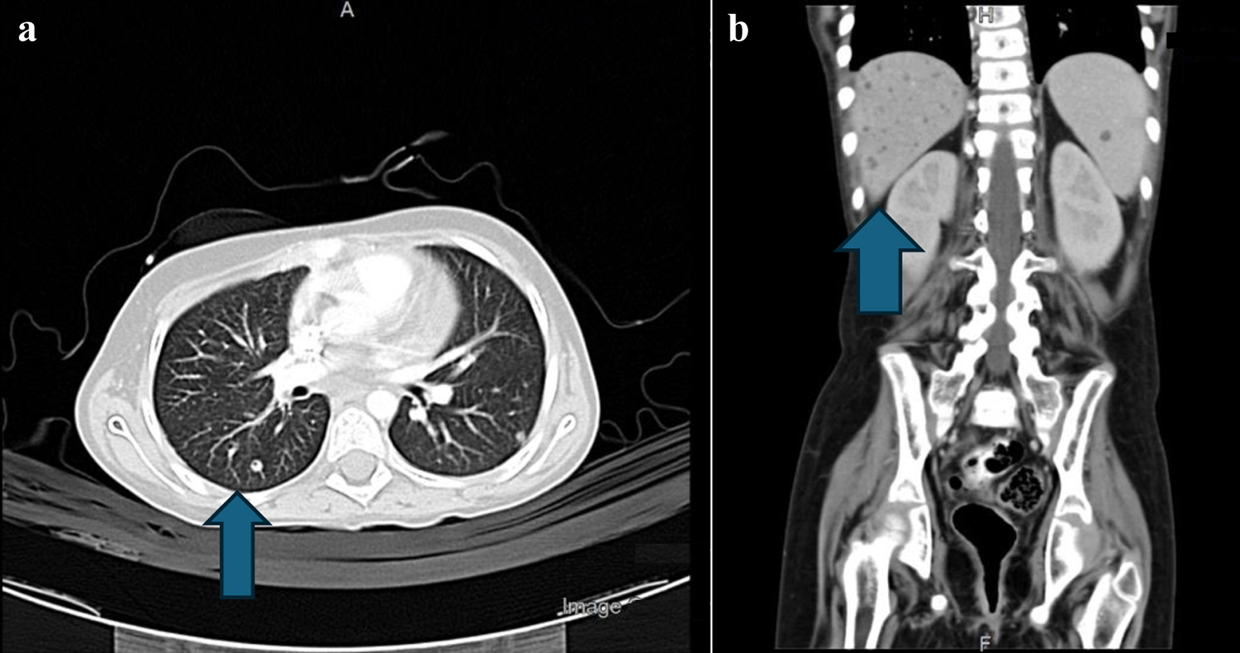
Journal of Medical Cases, ISSN 1923-4155 print, 1923-4163 online, Open Access
Article copyright, the authors; Journal compilation copyright, J Med Cases and Elmer Press Inc
Journal website https://jmc.elmerpub.com

Case Report

Volume 16, Number 5, May 2025, pages 174-180


Treatment of Invasive Fungal Disease During Therapy for Acute Lymphoblastic Leukemia

Figures

Figure 1.
Figure 1. (a) CT scan of the chest. The arrow points to right lung lower lobe, lateral basal segment small, tiny nodule. (b) Abdominal CT with contrast. The arrow shows numerous nodules in the liver, spleen, and kidneys, specifically pointing to right lower liver lobe small nodule. CT: computed tomography.
Figure 2.
Figure 2. (a) Ultrasound-guided biopsy of lung nodules showing hyphae with septate (possible Aspergillus). (b) Biopsy of lung nodules after fungal treatment.
Figure 3.
Figure 3. (a) CT scan of the chest. The arrow points to left lung lower lobe, lateral basal segment small, tiny nodule. (b) Abdominal CT with contrast. The arrow points to multiple nodules in the spleen. CT: computed tomography.