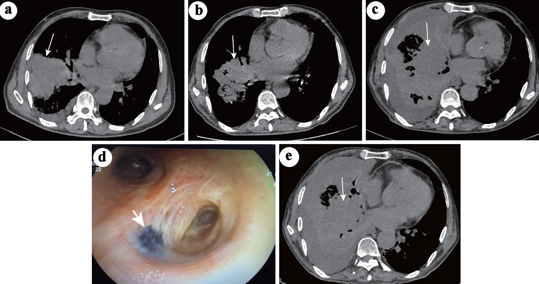
↓ Figure 1. Chest CT changes during the course of
pneumonia. (a) Large areas of consolidation (white arrow) in the middle-lower right lobe and pleural
effusion during the first hospitalization when the patient was infected with Mycoplasma
pneumoniae. (b) On the second admission, the consolidation (white arrow) in the right
middle-lower lobe was significantly absorbed and decreased, and the pleural effusion disappeared. (c)
After 2 weeks of nonspecific antifungal treatment, the consolidation (white arrow) in the right lobe
progressed, accompanied by obstructive inflammation and lymphangitis. Pleural effusion also appeared.
(d) Video bronchoscopy image showing a black fungating mass in the right principal bronchus (white
arrow). (e) No further progression in the right lobe (white arrow) after 26 days of anti-mucor therapy.
CT: computed tomography.