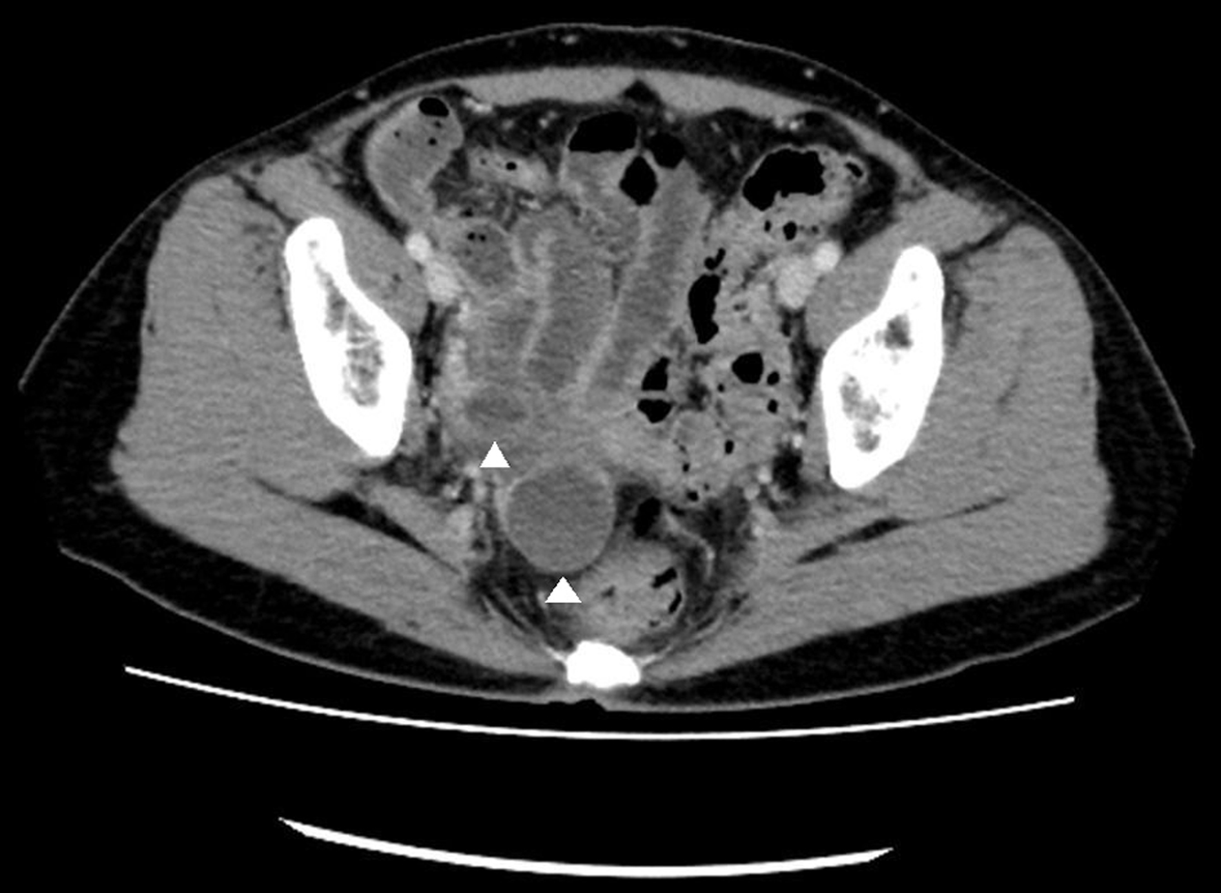
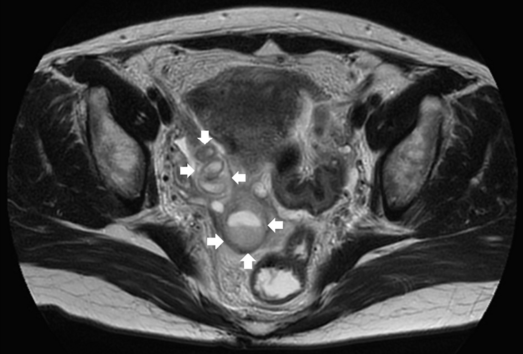
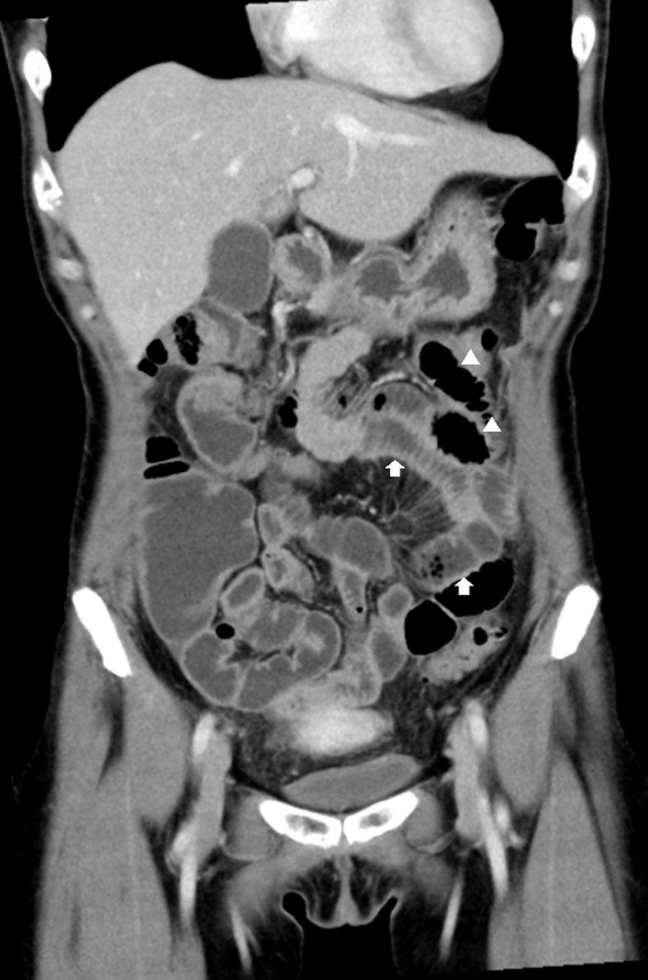
↓ Figure 1. Contrast-enhanced computed tomography
of the upper abdomen shows circular folds of Kerckring, thickening of the bowel wall (arrows), and small
intestinal loops containing gas (arrowheads).
| Journal of Medical Cases, ISSN 1923-4155 print, 1923-4163 online, Open Access |
| Article copyright, the authors; Journal compilation copyright, J Med Cases and Elmer Press Inc |
| Journal website https://jmc.elmerpub.com |
Case Report
Volume 16, Number 1, January 2025, pages 37-42
Peritonitis After Endometrial Cytology in a Woman With Hydrosalpinx Caused by Chronic Chlamydia trachomatis Infection
Figures