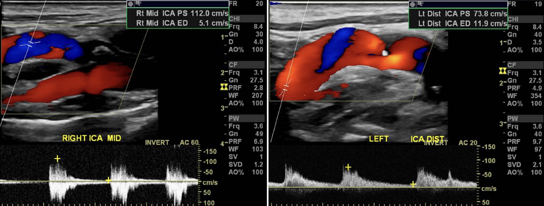
Journal of Medical Cases, ISSN 1923-4155 print, 1923-4163 online, Open Access
Article copyright, the authors; Journal compilation copyright, J Med Cases and Elmer Press Inc
Journal website https://jmc.elmerpub.com

Case Report

Volume 16, Number 2, February 2025, pages 82-86


Long-Term Outcomes and Management of Atypical Carotid Web in Nonagenarian

Figures

Figure 1.
Figure 1. Carotid duplex ultrasound of left and right ICA showing filling defects and stenosis. ICA: internal carotid artery.
Figure 2.
Figure 2. Coronal, sagittal, and axial views highlighting the right common and external carotid artery segments appear normal. There is approximately 80% stenosis of the proximal right internal carotid artery (white arrow). There is hypo-enhancement of the distal right internal carotid artery (black arrow).
Figure 3.
Figure 3. Intraoperative photo of excised section of right proximal internal carotid artery with the presence of a fibrous band protruding from the posterior lumen. Note the absence of an atheroma characteristic of atherosclerosis.
Figure 4.
Figure 4. Intraoperative photo highlighting the pin-hole opening rolled up and recreating natural intraluminal morphology (arrow).