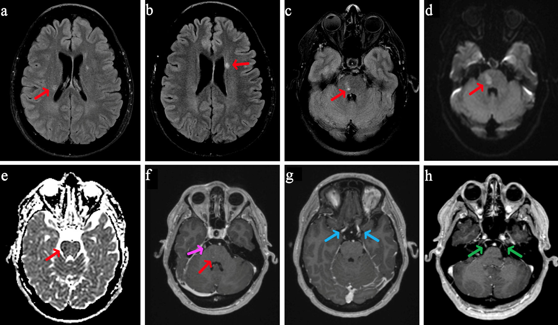
Journal of Medical Cases, ISSN 1923-4155 print, 1923-4163 online, Open Access
Article copyright, the authors; Journal compilation copyright, J Med Cases and Elmer Press Inc
Journal website https://jmc.elmerpub.com/

Case Report

Volume 15, Number 12, December 2024, pages 387-395


Exploring Overlap Syndromes: An Atypical Case of Multiple Sclerosis With Anti-Sjogren’s Syndrome Type B Antibody

Figures

Figure 1.
Figure 1. MRI brain with and without contrast. On axial T2-weighted FLAIR, there are two small periventricular white matter intensities along the bodies of the lateral ventricles (a, b, red arrows) and within the right para-midline pons (c, red arrow). On axial DWI (d), there is a small area of mild restricted diffusion in the right para-midline pons, measuring 1.4 × 1.0 cm (red arrow), with corresponding signal (red arrow) on ADC (e) and contrast enhancement on post-contrast MPRAGE (f, red arrow). Post-contrast MPRAGE imaging also shows bilateral enhancement of cranial nerves III (g, blue arrows), V (h, green arrows), and VI (f, pink arrow, unilateral only shown). MRI: magnetic resonance imaging; FLAIR: fluid-attenuated inversion recovery; DWI: diffusion-weighted imaging; ADC: apparent diffusion coefficient; MPRAGE: magnetization-prepared rapid gradient echo.
Figure 2.
Figure 2. MRI lumbar spine with contrast. On sagittal (a) and axial (b, d) T1-weighted post-contrast images, there is subtle enhancement of the cauda equina nerve roots (red arrows), with corresponding T2 axial (c, e) views. The upper dashed line on panel A corresponds to axial slices (b) and (c), whereas the lower dashed line corresponds to axial slices (d) and (e). MRI: magnetic resonance imaging.
Figure 3.
Figure 3. MRI thoracic spine with contrast. Axial (a) and sagittal (b) T1 MRI post-contrast images of the thoracic spine show subtle enhancement of the thoracic nerve roots (red arrows). MRI: magnetic resonance imaging.

Tables

Table 1. Serum and CSF Evaluation
 
EvaluationsValue (normal range)
aAbnormal results. Ig: immunoglobulin; Ab: antibody; CSF: cerebrospinal fluid; IFA: immunofluorescence assay; ANNA: anti-neuronal nuclear antibody; PCA: Purkinje cell antibody; DNER: delta/notch-like EGF-related receptor; AGNA/SOX1: anti-glial nuclear antibody/anti-SRY (sex determining region Y)-box 1; CRMP5/CV2: collapsin response mediator protein 5 (also known as “CV2”); MPO: myeloperoxidase; PR3: proteinase 3; C-ANCA: cytoplasmic antineutrophil cytoplasmic antibodies; P-ANCA: perinuclear antineutrophil cytoplasmic antibodies; MOG: myelin oligodendrocyte glycoprotein; SSA: Sjogren’s syndrome type A (also known as anti-Ro); SSB: Sjogren’s syndrome type B (also known as anti-La); CCP: cyclic citrullinated peptide; Rnp: ribonucleoprotein; CSF: cerebrospinal fluid; RBC: red blood cell; WBC: white blood cell; VDRL: venereal disease research laboratory test; GAD65: glutamic acid decarboxylase 65; AI: antibody index; EU: endotoxin units; APL-U: IgA phospholipid units; GPL-U: IgG phospholipid units; MPL-U: IgM phospholipid units.
Serum evaluation
  IgA level259.45 (84.5 - 499 mg/dL)
  IgG level764.35 (610.3 - 1,616 mg/dL)
  IgM level155.25 (35 - 242 mg/dL)
  Tissue IFA observationNo fluorescence observed
  ANNA1 (HU) Ab, IFANegative
  ANN2 (R1) Ab, IFANegative
  ANNA3 Ab, IFANegative
  PCA1 (YO) Ab, IFANegative
  PCA2 Ab, IFANegative
  PCA TR (DNER) Ab, IFANegative
  AGNA/SOX1 Ab, IFANegative
  AMPHIPHYSIN Ab, IFANegative
  CRMP5/CV2 Ab, IFANegative
  Aquaporin 4 Ab (IgG)Negative
  Anti-MPO Ab< 0.2 (0.0 - 0.9 U)
  Anti-PR3 Ab< 0.2 (0.0 - 0.9 U)
  C-ANCA< 1:20
  P-ANCA< 1:20
  Atypical P-ANCA< 1:20
  Lyme total AbNegative
  MOG Ab, serumNegative
  Sjogren’s Ab (SSA)< 0.2 (0.0 - 0.9 AI)
  Sjogren’s Ab (SSB)2.7, 2.8 (0.0 - 0.9 AI)a
  Ribosomal P Ab< 0.2 (0.0 - 0.9 AI)
  Salivary protein 1 IgG Ab2.6 (< 20 EU/mL)
  Salivary protein 1 IgA Ab2.0 (< 20 EU/mL)
  Salivary protein 1 IgM Ab74.2 (< 20 EU/mL)a
  Carbonic anhydrase VI IgG Ab> 160.0 (< 20 EU/mL)a
  Carbonic anhydrase VI IgA Ab87.0 (< 20 EU/mL)a
  Carbonic anhydrase VI IgM Ab17.2 (< 20 EU/mL)
  Parotid specific protein IgG Ab18.2 (< 20 EU/mL)
  Parotid specific protein IgA Ab27.4 (< 20 EU/mL)a
  Parotid specific protein IgM Ab22.0 (< 20 EU/mL; borderline: 20 - 25 EU/mL)
  Cyclic citrullinated peptide (CCP) Ab IgG< 16 (< 20)
  Lupus anticoagulantNot detected
  Rheumatoid factor< 14 (< 14 IU/mL)
  Cardiolipin Ab, IgA< 2.0 (< 20 APL-U/mL)
  Cardiolipin Ab, IgG< 2.0 (< 20 GPL-U/mL)
  Cardiolipin Ab, IgM< 2.0 (< 20 MPL-U/mL)
  Beta-2 glycoprotein I Ab, IgM< 2.0 (< 20 U/mL)
  Beta-2 glycoprotein I Ab, IgA< 2.0 (< 20 U/mL)
  Beta-2 glycoprotein I Ab, IgG< 2.0 (< 20 U/mL)
  Antinuclear Ab, IFANegative
  Anti-DNA (Ds) Ab3 (0-9 IU/mL)
  Rnp Ab< 0.2 (0.0 - 0.9 AI)
  Smith Ab< 0.2 (0.0 - 0.9 AI)
  Scleroderma (Scl-70) Ab< 0.2 (0.0 - 0.9 AI)
  Oligoclonal bands, serum0
CSF evaluationValue (normal range)
  Protein24 (15 - 45 mg/dL)
  Glucose71 (40 - 75 mg/dL)
  RBC count3,100 (0/mm3)a
  WBC count8 (0 - 5/mm3)a
  Neutrophils34%
  Lymphocytes59%
  Monocytes6%
  Eosinophils1%
  Cryptococcus gattii neoformans DNA CSFNot detected
  Cytomegalovirus DNA CSFNot detected
  Enterovirus RNA CSFNot detected
  Escherichia coli K1 DNA CSFNot detected
  Haemophilus influenzae DNA CSFNot detected
  Herpes simplex virus 1 DNA CSFNot detected
  Herpes simplex virus 2 DNA CSFNot detected
  Herpes virus 6 DNA CSFNot detected
  Parechovirus A RNA CSFNot detected
  Listeria monocytogenes DNA CSFNot detected
  Neisseria meningitidis DNA CSFNot detected
  Streptococcus agalactiae DNA CSFNot detected
  Streptococcus pneumoniae DNA CSFNot detected
  Varicella zoster virus DNA CSFNot detected
  VDRL, CSFNon-reactive
  CRMP5/CV2 AB, IFA, CSFNegative
  GAD65 antibody, IFA, CSFNegative
  Aquaporin 4 Ab (IgG), CSFNegative
  MOG Ab, CSFNegative
  Oligoclonal bands, CSF5 CSF-specific bands
  Flow cytometry analysisNo immunophenotypically abnormal T or B cells detected
  CytologyNegative for malignancy


Table 2. Longitudinal Disease Course
 
Time pointClinical findingsLaboratory dataImaging findingsTreatment
CSF: cerebrospinal fluid; MRI: magnetic resonance imaging; FLAIR: fluid-attenuated inversion recovery; IV: intravenous; SSB: Sjogren’s syndrome B.
Hospital admission (initial presentation)Left face and hemibody paresthesiasCSF: mild leukocytosis, 5 CSF-specific oligoclonal bandsMRI brain: T2 FLAIR hyperintensity in the bilateral periventricular regions and right pons; cranial nerve enhancement5-day course of high-dose IV methylprednisolone
Abnormal gait with left leg draggingSerum: positive anti-SSB antibody; no serum oligoclonal bandsMRI spine: enhancement in the cauda equina and thoracic nerve roots
Weakness in left hip and knee
Absent lower extremity reflexes
Hospital discharge (8 days post-admission)Mild improvement in strengthNo new laboratory findingsNo new imaging findingsDischarged with follow-up to neuroimmunology and rheumatology
Persistent left-sided paresthesias
2 months post-dischargeImproved motor examSerum: elevated salivary protein 1 IgM, elevated carbonic anhydrase VI IgG and IgM antibodies, elevated parotid specific protein IgA antibodyMRI brain: persistent enhancement of cranial nerves3-day course of high-dose oral prednisone
Residual numbnessOfatumumab 20 mg monthly


Table 3. A Case-Based Literature Review of MS and SS Overlaps and Diagnostic Dilemmas
 
CasePatientCase presentationSuspected initial diagnosisDiagnostic dilemmas and changesFinal diagnosisKey takeaways
F: female; BLE: bilateral lower extremity; MS: multiple sclerosis; SSB: Sjogren’s syndrome B; SS: Sjogren’s syndrome; MRI: magnetic resonance imaging; CNS: central nervous system; UCTD: undifferentiated connective tissue disease; IFN: interferon; PBC: primary biliary cholangitis; BUE: bilateral upper extremity; N/A: not available.
Our caseF, 39 yearsPatient presented with left face and hemibody paresthesias, abnormal gait with left leg dragging, weakness in left hip and knee, and absent BLE reflexes.MSSerum studies revealed a positive Sjogren’s anti-SSB antibody, despite no history of systemic or sicca symptoms.MS and SS overlapIt is important to consider autoimmune overlap conditions in cases where there are atypical neurologic symptoms, as the co-occurrence of MS and SS can impact treatment strategies.
Mohamednour et al, 2020 [10]F, 58 yearsPatient with a 3-year history of MS presented with polyarthralgia, sicca symptoms, and Raynaud’s.MSInitial MRI of the brain and spine were reported as normal with few “nonspecific white matter areas”, complicating the diagnosis.SSCNS involvement in SS can closely resemble MS, complicating the diagnosis. Peripheral neuropathy and vasculitis are key differentiators. Early neurophysiological testing is crucial, and effective immunosuppression with shared decision-making can achieve successful outcomes.
Jung et al, 2000 [11]F, 45 yearsPatient with a 4-year history of dry eyes and mouth presented with BLE weakness, voiding difficulty, decreased visual acuity, BLE spasticity and hyperesthesia, and diffuse hyperreflexia.MSErythematous skin lesions soon developed in the periauricular area and BLE. Skin and salivary gland biopsies both showed lymphocytic infiltration.SSSS should be considered as a differential diagnosis of a patient presenting with a syndrome that resembles MS because of the similarity in clinical manifestations and laboratory findings of both diseases.
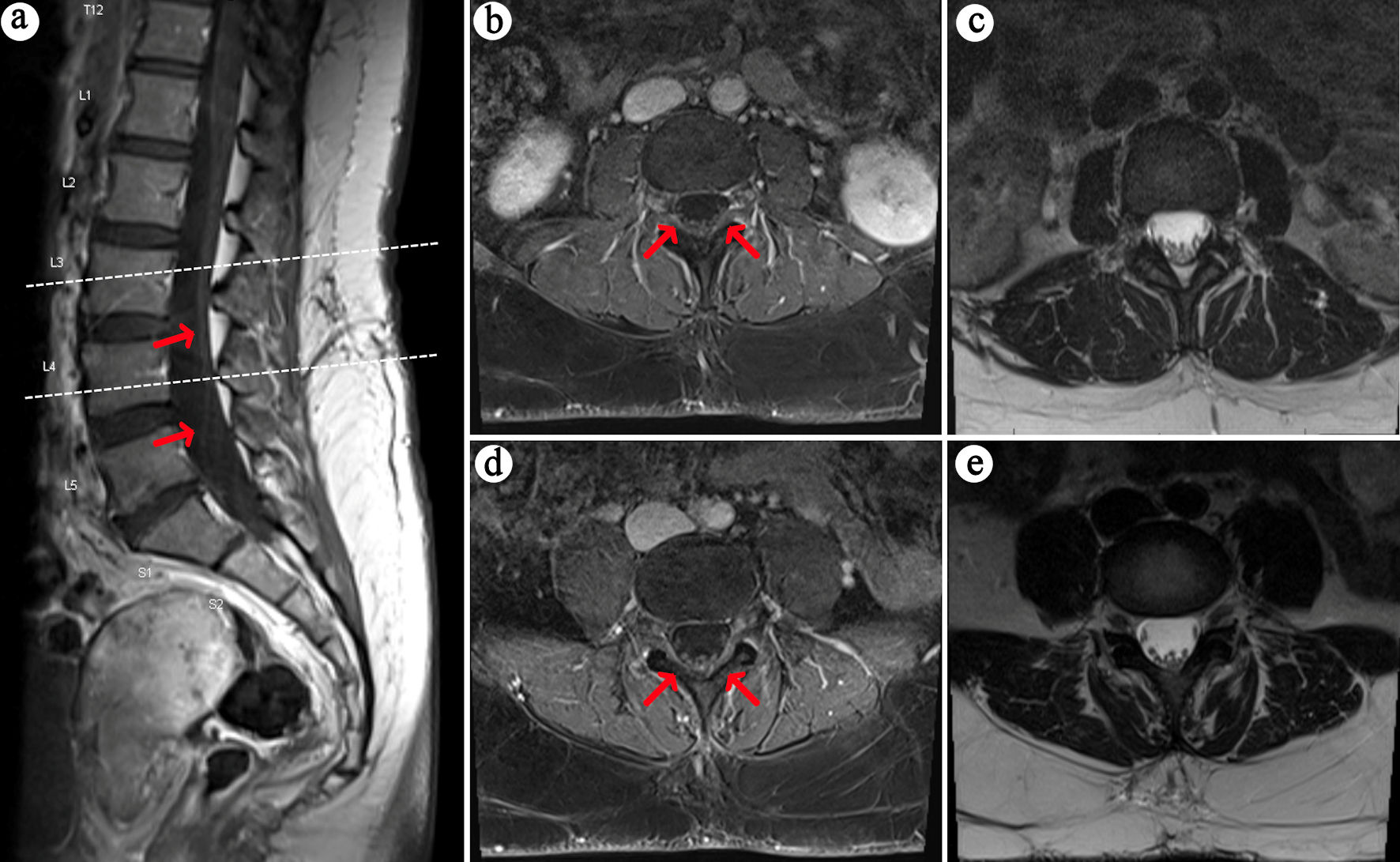
Thong et al, 2002 [12]F, 40 yearsPatient with a history of a lacunar stroke with no residual deficits presented with acute urinary retention and diminished sensation below T5 spinal level, followed by a year of episodes of ataxia and upper limb dysmetria, retrobulbar neuritis, and pseudobulbar palsy.UCTDFive years after initial neurologic symptoms, the patient presented with pseudobulbar palsy and endorsed 1 year of sicca symptoms. Schirmer test was positive, and anti-Ro antibody was elevated.SSPatients with MS-like diseases should be carefully evaluated for other system autoimmune conditions as the treatment and prognosis differ from MS. Particularly, sicca symptoms should be sought as they often go unnoticed by patients.
De Santi et al, 2005 [13]F, 48 yearsPatient with a 29-year history of MS treated well with IFN-beta 1a for 5 years presented with xerophthalmia and xerostomia with dysphagia.MSN/AMS and SS overlapSS can develop long after the onset of MS, even during successful IFN-beta treatment. This case highlights the need to monitor MS patients for SS, as symptoms may arise unexpectedly, complicating diagnosis and management.
Guzel et al, 2022 [14]F, 51 yearsPatient with a history of PBC and Hashimoto thyroiditis presented with numbness and weakness in the left upper and lower extremities; further questioning revealed symptoms of dry eyes and mouth.MS and SS overlapN/AMS and SS overlapThis case highlights the rare coexistence of four autoimmune diseases, emphasizing the importance of considering multiple overlapping autoimmune disorders in complex presentations.
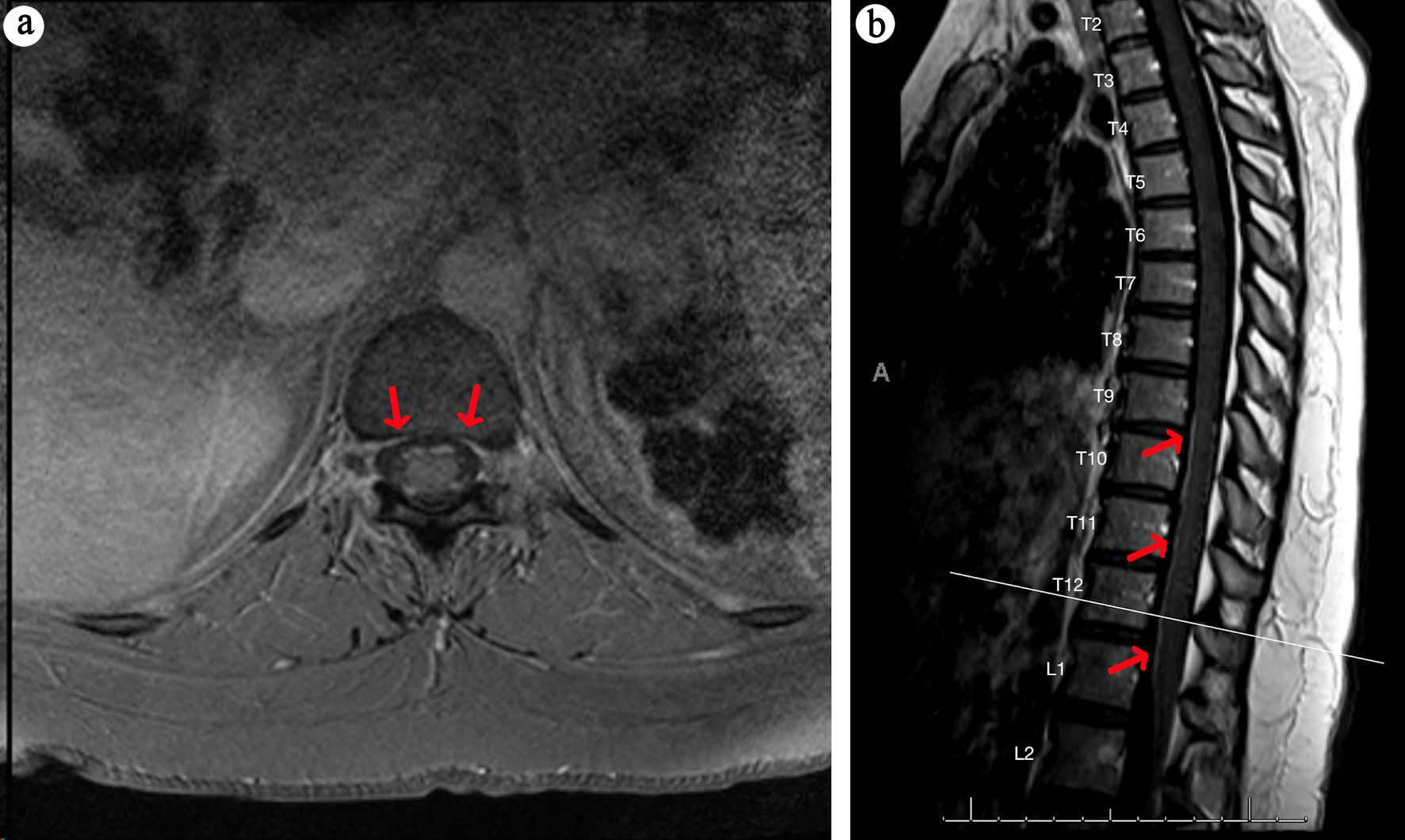
Liu et al, 2014 [15]F, 75 yearsPatient presented with recurring numbness and sensory changes in BUE and BLE with zonesthesia in the waist.MSPatient had subsequent worsening of paresthesias and swelling of the parotid gland; oral and ocular sicca syndromes were suspected from history taking and confirmed by Schirmer test and labial salivary gland biopsiesSSSS should be considered in the differential diagnosis of patients presenting with MS-like neurological symptoms, as it can mimic MS both clinically and neuroradiologically.