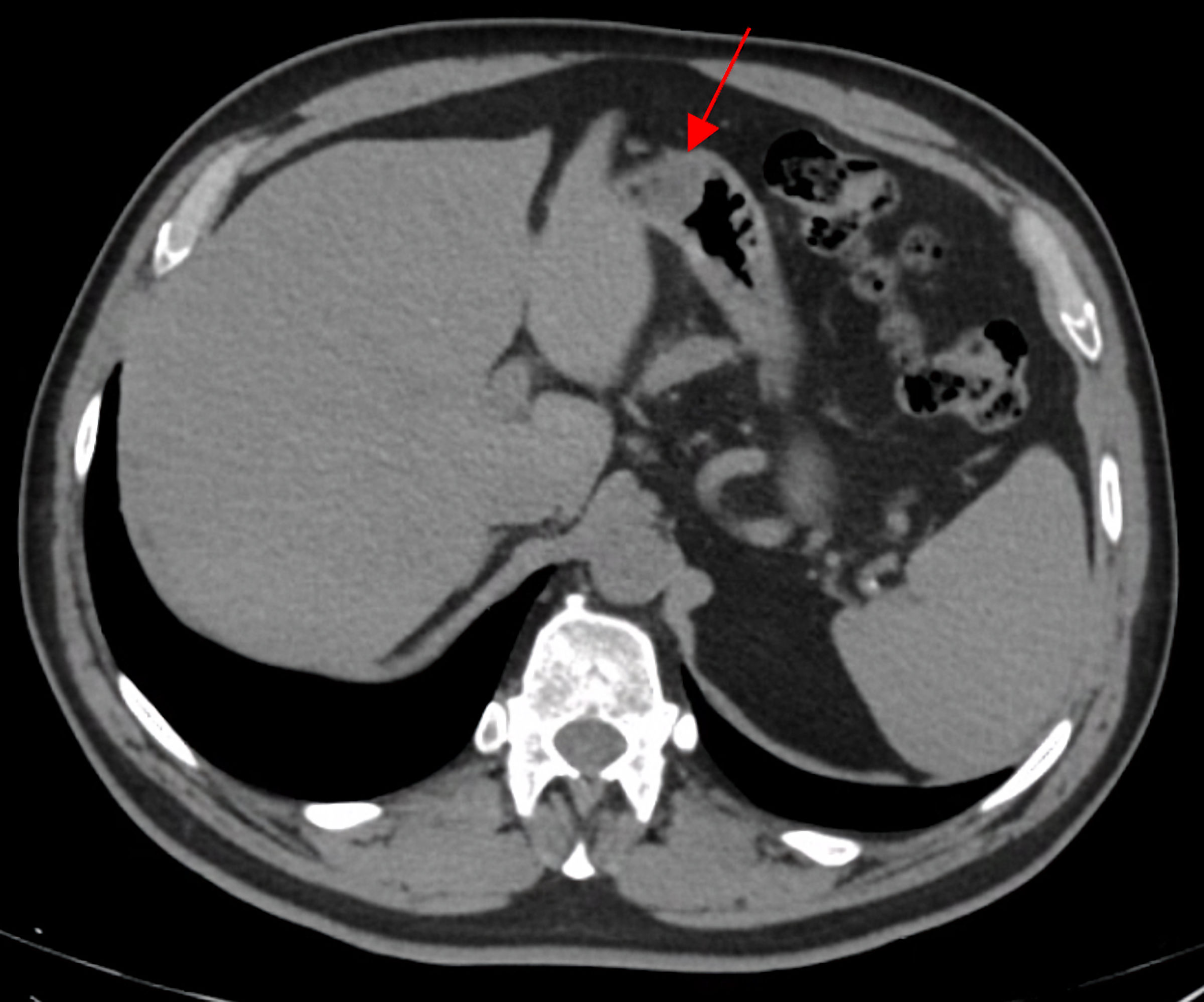
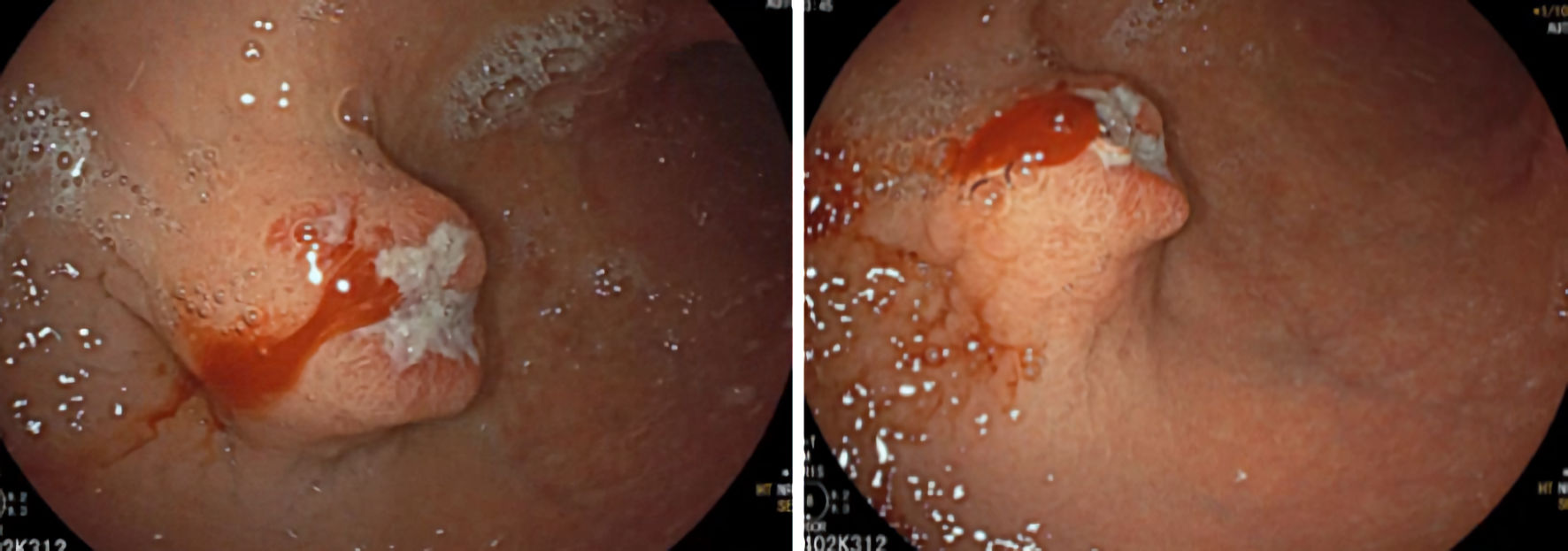
Journal of Medical Cases, ISSN 1923-4155 print, 1923-4163 online, Open Access
Article copyright, the authors; Journal compilation copyright, J Med Cases and Elmer Press Inc
Journal website https://jmc.elmerpub.com/

Case Report

Volume 15, Number 12, December 2024, pages 371-375


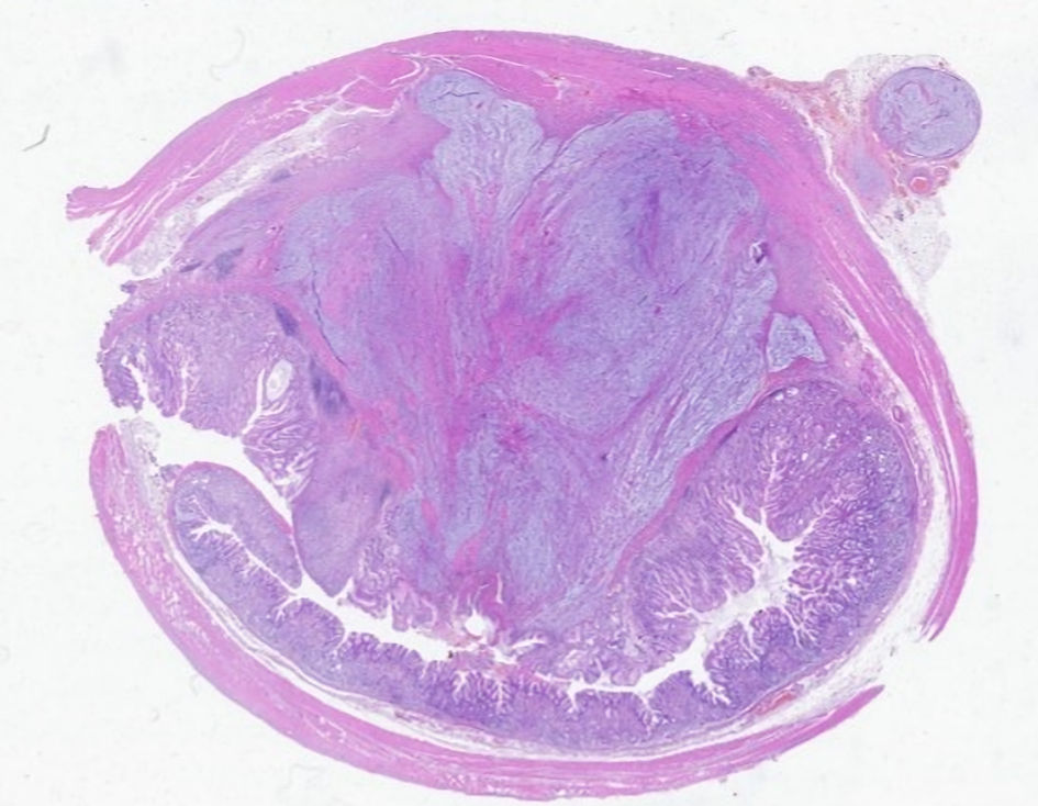
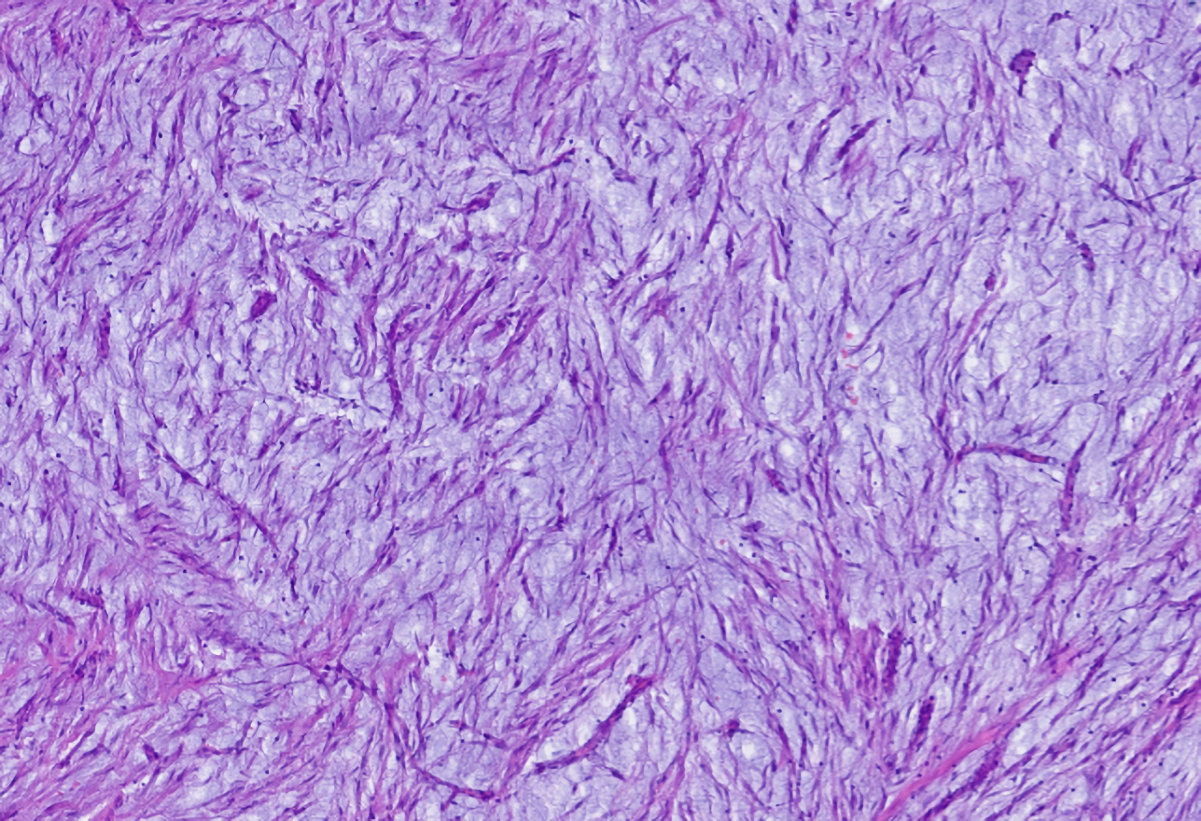
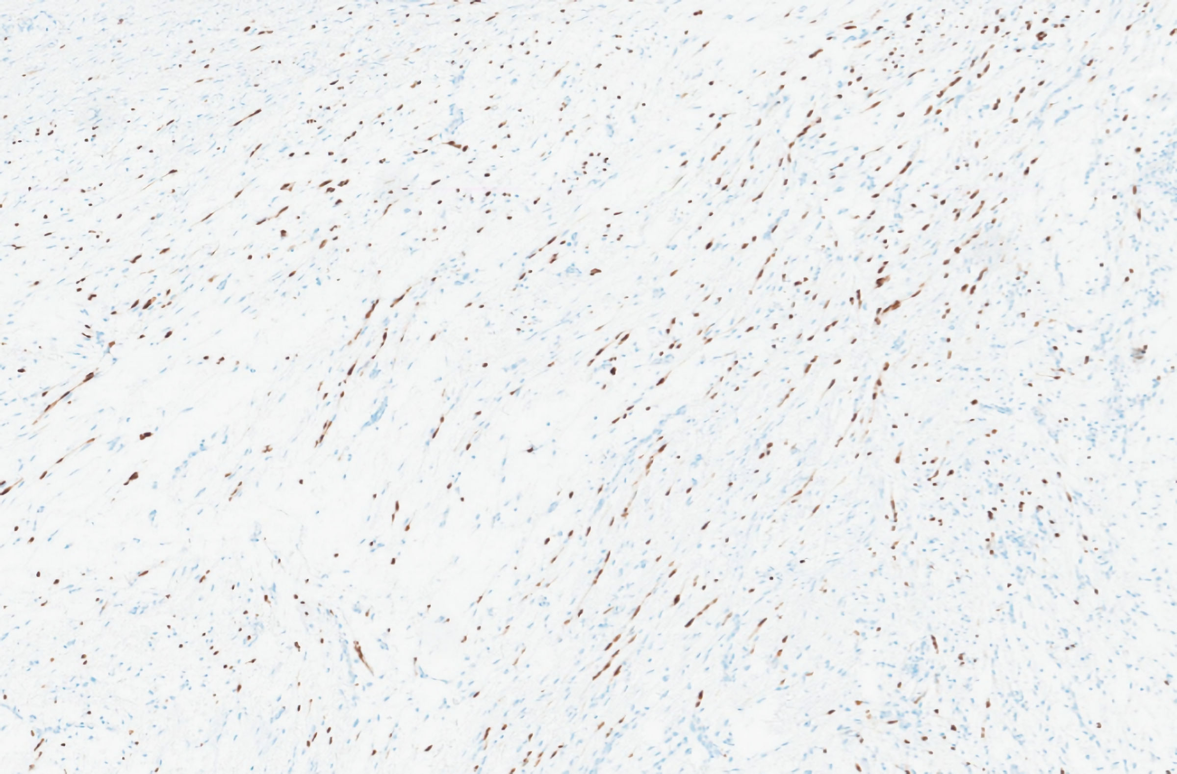
Gastric Schwannoma: A Rare Cause of Gastric Bleeding

Figures

Figure 1.
Figure 1. Upper gastrointestinal endoscopy showing subepithelial lesion located on the anterior surface/greater curvature of the middle/distal body, ulcerated.
Figure 2.
Figure 2. Abdominal and pelvic tomography scan. In a submucosal position, on the anterior wall of the transition between the body and the gastric antrum, a well-defined hypodense solid nodularity, with homogeneous and frank enhancement, measuring 22 × 18 mm (red arrow) was observed.
Figure 3.
Figure 3. Microscopic analysis. The lesion developed in dependence on the gastric wall.
Figure 4.
Figure 4. Microscopic analysis showing proliferation of spindle cells on a background with a myxoid appearance.
Figure 5.
Figure 5. Immunohistochemical analysis showing SOX10 immunoreactive cells.