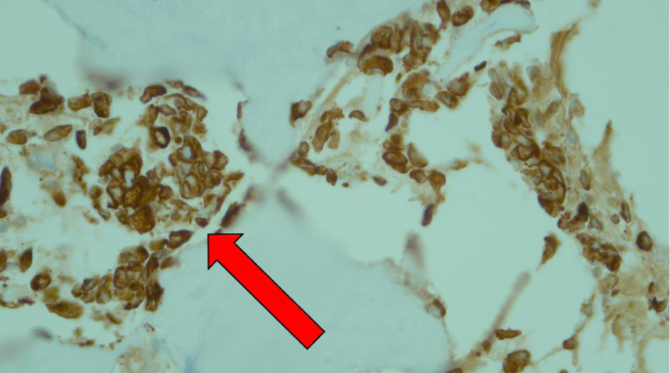
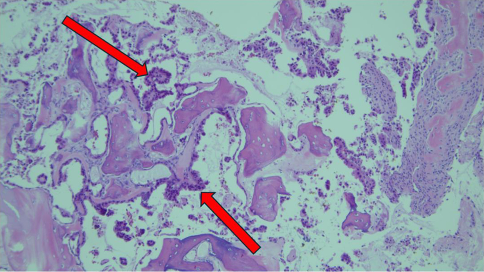
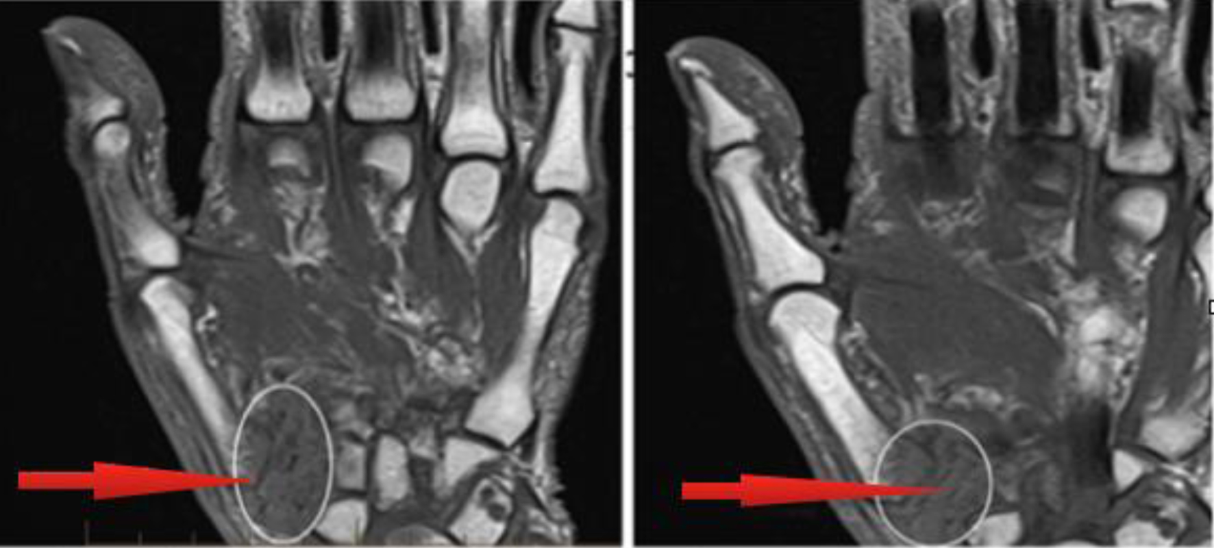
Journal of Medical Cases, ISSN 1923-4155 print, 1923-4163 online, Open Access
Article copyright, the authors; Journal compilation copyright, J Med Cases and Elmer Press Inc
Journal website https://jmc.elmerpub.com

Case Report

Volume 16, Number 4, April 2025, pages 131-134


An Unusual Case of Pancreatic Adenocarcinoma Metastasis to the Trapezium Bone

Figures

Figure 1.
Figure 1. Metastatic involvement of entire trapezium bone and base of first metacarpal associated with diffuse edema of soft tissue, with red arrows indicating metastatic involvement of pancreatic carcinoma.
Figure 2.
Figure 2. Hematoxylin stain from surgical biopsy of left trapezium with red arrow depicting a nest of atypical cells supporting suspicion for malignant pancreatic carcinoma cells.
Figure 3.
Figure 3. Immunohistochemistry staining from surgical biopsy of left trapezium. Red arrows depict strong cytoplasmic staining depicting neoplastic cells that are pancytokeratin (+), with CK7 (variably +), CK20 (-), CDX2 (-), and TTF1 (-). The neoplastic cells were noted with high expression of CD68, highlighting abundant histocytes. These findings were consistent with metastatic pancreatic adenocarcinoma.