| Journal of Medical Cases, ISSN 1923-4155 print, 1923-4163 online, Open Access |
| Article copyright, the authors; Journal compilation copyright, J Med Cases and Elmer Press Inc |
| Journal website https://jmc.elmerpub.com |
Case Report
Volume 18, 2027, e5449

A Rare Clinical Case of Thyrotoxic Periodic Hypokalemic Paralysis With Unilateral Lower-Limb Weakness
Assel Meiramovaa, b  , Yerzhan Khochshanovb , Zhibek Dautbayevaa , Viktoriya Nazarovaa, b , Zhanar Akhmetovaa, c, d , Yelvira Omralinab, d , Bayan Ainabekovab
aScientific Department, Art Science Innovation Center, Astana, Kazakhstan
bDepartment of Internal Medicine No. 2, NJSC Astana Medical University, Astana, Kazakhstan
cDepartment of Rehabilitation and Sports Medicine, NJSC Astana Medical University, Astana, Kazakhstan
dCorresponding Authors: Zhanar Akhmetova, Department of Rehabilitation and Sports Medicine, Astana Medical University, Astana 010000, Kazakhstan; Yelvira Omralina, Department of Internal Medicine No. 2, NJSC Astana Medical University, Astana, Kazakhstan
Manuscript submitted October 7, 2026, accepted November 4, 2026, published online November 20, 2026
Short title: TPP With Unilateral Lower-Limb Weakness
doi: https://doi.org/10.14740/jmc5449
| Abstract | ▴Top |
Thyrotoxic hypokalemic periodic paralysis (TPP) is a rare complication of thyrotoxicosis characterized by transient episodes of muscle weakness associated with hypokalemia. The clinical presentation typically includes symmetrical proximal weakness of the lower extremities. Atypical manifestations, especially those mimicking focal neurological disorders, may lead to delayed diagnosis. We describe a case of TPP presenting with recurrent asymmetric muscle weakness, manifesting as transient unilateral lower-limb weakness in a male patient. The episodes occurred predominantly at night and resolved spontaneously without treatment. Due to the unilateral nature of the motor deficit, the condition was initially misinterpreted as transient ischemic attacks, which led to a diagnostic delay of approximately 1 year. Neuroimaging revealed only nonspecific stable white matter lesions, and neurological therapy did not result in clinical improvement. Subsequent endocrinological evaluation demonstrated overt thyrotoxicosis, with elevated free thyroid hormone levels and high titers of thyroid-stimulating hormone receptor antibodies (TRAb), consistent with Graves’ disease. Initiation of antithyroid therapy and beta-blocker treatment led to normalization of thyroid hormone levels and complete resolution of the episodes of muscle weakness. This case illustrates a rare atypical presentation of TPP, manifesting as unilateral lower-limb weakness and mimicking a transient ischemic attack. The report highlights the importance of considering thyrotoxicosis in the differential diagnosis of transient focal neurological deficits, especially in patients with recurrent reversible episodes of muscle weakness.
Keywords: Thyrotoxic hypokalemic periodic paralysis; Diffuse toxic goiter; Muscle weakness; Transient ischemic attack
| Introduction | ▴Top |
Clinical presentation of the diffuse toxic goiter is determined by excessive production of thyroid hormones, resulting in thyrotoxicosis of varying severity. However, rare atypical variants of disease onset have been described in which the condition manifests not with the predominant typical symptoms of thyrotoxicosis and/or ophthalmopathy, but with skeletal muscle involvement in the form of thyrotoxic hypokalemic periodic paralysis (TPP). Such observations emphasize that typical thyrotoxic signs may be minimal or absent, while the clinical picture is characterized by episodic muscle weakness in the setting of biochemical thyrotoxicosis and hypokalemia, which may lead to diagnostic errors and delays in etiological treatment [1]. According to available data, TPP most commonly occurs in men, predominantly of Asian origin, and typically develops between the ages of 20 and 40 years. In many reported cases, patients seek emergency care at night or in the morning after awakening. This pattern is associated with circadian changes in hormonal and insulin responsiveness, as well as typical triggers such as evening exertion or a large dinner, followed by a nocturnal attack and a morning visit to the hospital [2].
We present a rare case of unilateral lower-limb weakness in a male patient with thyrotoxic periodic paralysis, in whom the diagnosis was delayed by 1 year. The clinical presentation mimicked a transient ischemic attack (TIA) or stroke. This case report was prepared in accordance with the CARE guidelines.
| Case Report | ▴Top |
In November 2025, a 50-year-old man of Kazakh ethnicity presented to an endocrinologist with complaints of truncal hyperhidrosis, palpitations on minimal exertion, intermittent weakness in the muscles of the lower limb muscles, and a 10-kg weight loss over the preceding year.
According to the patient’s history, the first episode occurred 1 year earlier, on November 7, 2024. While in his usual state of good health, one morning after a routine breakfast, he suddenly experienced weakness in his lower limbs, particularly on the left side. He was unable to remain standing and fell. Colleagues transported him to an outpatient clinic, where he was examined by a neurologist and subsequently urgently transferred by emergency medical services to the emergency department of a multidisciplinary city hospital. On admission, the patient complained of numbness in the left limbs, weakness in the left lower limb, and pain in the anterior thigh muscles. Physical examination revealed pale skin. Vital signs were as follows: body temperature 36.7 °C, respiratory rate 16 breaths per minute, blood pressure 125/80 mm Hg, heart rate 74 bpm, and SpO2 97%. The ABCD2 score was 5 points, indicating moderate risk of stroke. Neurological examination demonstrated clear consciousness (Glasgow Coma Scale score of 15), negative meningeal signs, and intact cranial nerves (no oculomotor deficits, symmetrical facial movements, and preserved pharyngeal reflexes). Motor examination showed normal muscle tone and full strength in all limbs (5/5). Tendon reflexes were symmetric (D = S). The Barre test was negative. A mild intention tremor was observed during the finger-to-nose and heel-to-shin tests. No pathological reflexes were elicited. Sensory examination findings were normal. The patient was hospitalized with a diagnosis of G45.8 cerebrovascular disease, TIA (November 2024), presenting as transient unilateral leg weakness.
During the initial hospitalization, treatment was directed toward the presumed neurological diagnosis and included anticoagulant and antiplatelet therapy, infusion therapy, vasoactive and neuroprotective medications, as well as symptomatic treatment. No specific potassium replacement therapy was administered. Thyroid function was not assessed during this hospitalization. Blood pressure monitoring was recommended, with antihypertensive therapy as needed, along with follow-up by a neurologist at his place of residence.
After discharge from the hospital, the patient had repeated outpatient consultations with neurologists because of recurrent episodes of muscle weakness in the left lower limb. The weakness occurred predominantly at night and resolved spontaneously by morning or later the same day without medication. The patient reported difficulty changing body position, particularly when turning from his back onto his side, noting that he “had to move his leg with his hands to complete the turn.” The episodes occurred up to once or twice per month. Because of these recurrent attacks of muscle weakness, the patient was forced to stop working. According to the patient, while searching for the cause of his condition and experiencing significant anxiety, he noticed progressive weight loss. He was unable to specify the exact onset of associated symptoms such as increased sweating and palpitations. The prescribed neuroprotective therapy did not result in subjective clinical improvement.
Because of the current episodes of unilateral leg weakness and the absence of a clear neurological explanation, the patient was subsequently referred for endocrinological evaluation.
The patient had no history of chronic diseases, except for benign prostatic hyperplasia, and no family history of similar episodes. He denied any family history of endocrine disorders. He previously smoked one pack of cigarettes per day for 15 years but quit 4 years before the onset of symptoms.
Laboratory testing performed on the day of admission (November 7, 2024) demonstrated hypokalemia, with serum potassium concentration of 3.1 mmol/L (Table 1). Serum potassium was not remeasured during the investigation period; therefore, the timing and mechanism of normalization could not be determined. The next documented serum potassium measurement was obtained 1 year later, during the endocrinology evaluation in November 2025.
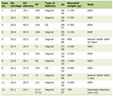 Click to view | Table 1. Laboratory Test Results Obtained During the Initial Hospitalization for Acute Unilateral Lower-Limb Weakness and Suspected TIA |
To evaluate acute cerebrovascular pathology, a non-contrast computed tomography (CT) scan of the brain was performed. No intracranial hemorrhage or acute ischemic changes were identified, and only chronic microangiopathic changes were noted. Electrocardiography (ECG), performed on November 8, demonstrated sinus tachycardia, with a heart rate of 95 beats per minute.
Brain magnetic resonance imaging (MRI) was subsequently performed to evaluate for a possible structural cause of the recurrent unilateral lower-limb weakness. MRI scans performed in November 2024 and August 2025 revealed a small number of vasogenic foci in the white matter of both hemispheres, without evidence of negative progression (Fig. 1). No structural lesion explaining the recurrent unilateral weakness was identified. Because of the recurrent episodes of unilateral leg weakness without a clear neurological explanation, the patient was subsequently referred for endocrinological evaluation.
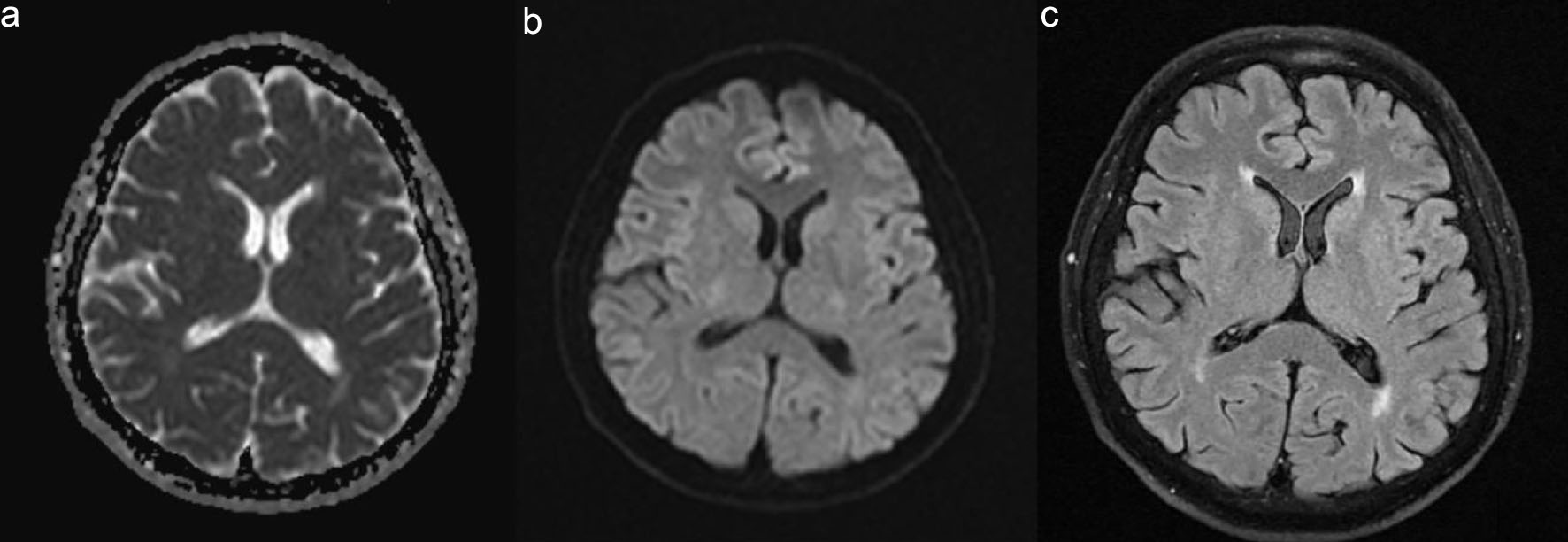 Click for large image | Figure 1. Brain MRI in a patient with unilateral muscle weakness mimicking transient ischemic attack. (a) Axial diffusion-weighted imaging (DWI) demonstrating no focal hyperintense lesions suggestive of acute ischemia, November 2024. (b) Axial apparent diffusion coefficient (ADC) map showing no diffusion restriction, November 2024. (c) Axial FLAIR image demonstrating small nonspecific white matter hyperintense lesions without radiological progression compared with the baseline MRI, August 2025. MRI: magnetic resonance imaging. |
Thyroid function was not assessed during the initial hospitalization or during subsequent neurological examinations. It was first evaluated during an endocrinology consultation in November 2025, approximately 1 year after the initial episode of weakness. At the time of examination by the endocrinologist: objectively, the skin was of normal color and moist, with hyperhidrosis of the trunk and palms, height was 163 cm, weight was 56 kg, and body mass index was 21.1 kg/m2. Palpation of the thyroid gland revealed slight enlargement, with each lateral lobe slightly larger than the terminal phalanx of the patient’s thumb, painless, with no abnormalities of the regional lymph nodes. A hand tremor was noted. The “telegraph pole” sign was negative, as was Pemberton’s sign. Ocular signs were absent. Cardiac activity was regular. Blood pressure was 127/76 mm Hg. Heart rate was 99 beats per minute. No pathological murmurs were detected. Neurological examination revealed no abnormalities. Findings in other organs and systems were unremarkable.
Laboratory evaluation in November 2025 demonstrated normal renal function (creatinine 67.2 µmol/L, reference range 44–97) and normal potassium level (3.9 mmol/L, reference range 3.5–5.1). The complete blood count was within normal limits. No abnormalities of carbohydrate metabolism were detected (HbA1c 5.44%, reference range 4.8–5.9). Thyroid function tests showed suppressed thyroid-stimulating hormone (TSH, < 0.001 µIU/mL, reference range 0.40–3.77), elevated free T3 (6.40 pg/mL, reference range 2.0–4.40) and free T4 (4.86 ng/dL, reference range 1.0–1.6), and markedly increased TSH receptor antibodies (64.1 IU/L, reference range less than 1.75), consistent with active Graves’ disease.
A diagnosis of grade 1 diffuse toxic goiter (according to WHO recommendations) was established, with moderate thyrotoxicosis. TPP was diagnosed.
Treatment with antithyroid medication was initiated: Thyrozol (thiamazole) 30 mg/day, and beta-blocker therapy with bisoprolol 5 mg/day.
At the 4-week follow-up, free T3 decreased from 6.40 to 3.50 pg/mL and free T4 from 4.86 to 0.72 ng/dL, while TSH remained suppressed (< 0.001 µIU/mL). The patient reported marked clinical improvement, and no further episodes of recurrent unilateral lower-limb weakness were reported following initiation of antithyroid and beta-blocker therapy. After the 4-week follow-up, the patient relocated and continued follow-up at another healthcare facility. According to the patient, his general condition remained satisfactory; however, subsequent clinical records and laboratory data were not available to our team. The absence of recurrent attacks following treatment of thyrotoxicosis was consistent with the proposed diagnosis of TPP, although this temporal association does not by itself establish causality.
| Discussion | ▴Top |
This clinical case illustrates recurrent, stereotyped episodes of isolated weakness in the left lower limb, prompting a broad differential diagnosis among conditions presenting with transient focal neurological deficits. Given the sudden onset and complete reversibility of symptoms, a TIA of either cerebral or spinal origin was initially considered. However, the recurrent stereotyped episodes over the following year, together with the absence of sensory, cortical, or pelvic disturbances, and the lack of a corresponding structural lesion on serial neuroimaging, made a cerebrovascular cause less likely [3]. A focal epileptic seizure with Todd’s paralysis was considered less likely on the overall clinical course because there were no reported clonic phenomena, altered consciousness, and a postictal period characteristic of the post-seizure phase [4]. Peripheral neurological causes, including radiculopathy or mononeuropathy, were considered less likely because of the recurrent, transient, and spontaneously reversible nature of episodes and the absence of persistent neurological deficits between attacks [5]. Metabolic and mitochondrial disorders were also considered. In particular, mitochondrial cytopathy (e.g., MELAS syndrome) was considered less likely on the overall clinical course because of absence of accompanying symptoms such as headache, vomiting, seizures, or lactic acidosis, together with the lack of characteristic MRI changes (lesions not corresponding to vascular territories) [6].
Taken together, the recurrent episodes of transient weakness and absence of a clear neurological explanation prompted consideration of a metabolic or neuromuscular cause. The detection of hypokalemia during the initial hospitalization for suspected TIA, a history of recurrent episodes of transient unilateral lower-limb weakness over the following year, and the subsequent identification of thyrotoxicosis approximately 1 year later supported the diagnosis of TPP.
This case corresponds to descriptions in the literature of TPP occurring in men, characterized by nocturnal attacks and the absence of an association with physical exertion [7]. Some authors have noted a seasonal pattern of clinical manifestations during the summer period, attributing this to increased perspiration in hot weather [1, 8, 9]. However, in our clinical case, this trend was not observed. Unlike previously reported cases, carbohydrate-rich dietary trigger could not be confirmed in our patient because no dietary record was available. Further supporting this diagnosis was the presence of sinus tachycardia on ECG recorded during hospitalization, followed by progressively worsening symptoms of hyperthyroidism. Possible signs of a prodromal period have also been described, including pain, muscle cramps, and stiffness in the affected muscles [1, 10]. In our patient, upon admission to the hospital, complaints of pain in the anterior thigh muscle were documented, which could not be explained within the framework of a TIA diagnosis.
However, the localization and asymmetric involvement observed in this clinical case fall outside the classical presentation of TPP, which ultimately led to a 1-year delay in establishing the correct diagnosis. Reported cases of TPP are predominantly characterized by the development of paraplegia or paraparesis, with preferential involvement of the proximal muscles of the lower extremities rather than the upper extremities. Symmetric muscle involvement is more commonly described, although cases of quadriparesis have also been reported [11–14]. Some authors have noted involvement of the pelvic girdle muscles in addition to the muscles of the lower limbs [15].
We identified one publication describing atypical manifestation in the form of asymmetric paralysis, in which a patient with a prior history of Graves’ disease experienced sudden unilateral weakness on the right side of the body [16]. Another report described atypical weakness beginning in the upper limbs, with asymmetric involvement between the two sides [10]. We were unable to find any published cases of TPP presenting as unilateral lower-limb weakness.
Following initiation of antithyroid and beta-blocker therapy, no further episodes of transient unilateral leg weakness were reported during follow-up, supporting the proposed diagnosis, although this temporal association does not establish causality.
The 1-year delay in establishing the diagnosis also deserves particular consideration. In retrospect, the hypokalemia documented during the initial hospitalization represented an important diagnostic clue. However, the unilateral nature of the lower-limb weakness prompted evaluation for an acute cerebrovascular event and may have shifted attention away from a metabolic cause of the motor deficit. Because serum potassium was not subsequently reassessed and thyroid function testing was not performed at the time, the underlying thyrotoxicosis remained unrecognized.
This case highlights the importance of considering metabolic and endocrine causes alongside vascular, epileptic, spinal, and mitochondrial causes in patients presenting with recurrent motor paroxysms. Thyrotoxic hypokalemic periodic paralysis, although rare and potentially treatable, may mimic stroke or TIA and should therefore be included in the differential diagnosis of unilateral neurological deficits, particularly when hypokalemia is present. An episode of paralysis may also represent the first clinical manifestation leading to the diagnosis of previously unrecognized thyrotoxicosis.
Learning points
1. TPP may present atypically as unilateral lower-limb weakness, mimicking focal neurological conditions such as TIA.
2. Recurrent, reversible episodes of muscle weakness, especially occurring at night, should prompt evaluation of serum potassium and thyroid function, even in the absence of classic bilateral symptoms.
3. Integrating neurological and endocrine findings in patients presenting with recurrent transient weakness may facilitate earlier recognition of thyrotoxicosis and reduce diagnostic delay.
Acknowledgments
The authors would like to thank the patient.
Financial Disclosure
This clinical case was described as part of the grant funded research project entitled “Gut-eye axis in diffusely toxic goiter: features of the microbiome of two econishes.” This research was funded by the Science Committee of the Ministry of Science and Higher Education of the Republic of Kazakhstan (Grant No. AP26100297).
Conflict of Interest
The authors declare no conflict of interest.
Informed Consent
Written informed consent was obtained from the patient for publication of this case report and any accompanying images. Ethical approval was obtained from local ethics committee of ArtScience Innovation Center LLP (Protocol No. 9-2024 of 10.10.2024).
Author Contributions
AM: conception and design of the study; AM and BA: endocrinological examination and interpretation of thyroid tests; ZD and YK: collection of patient history and clinical data; VN and YO: coordination of laboratory diagnostics; ZA: neurological examination neuroimaging interpretation; ZD: ophthalmological examination and assessment of orbitopathy; ZA, BA, and YO: drafting the initial manuscript and critical revision. All authors read and approved the final manuscript.
Data Availability
The data supporting the findings of this study are available from the corresponding author upon reasonable request.
Abbreviations
CT: computed tomography; ECG: electrocardiography; MRI: magnetic resonance imaging; TPP: thyrotoxic hypokalemic periodic paralysis; TSH: thyroid-stimulating hormone
| References | ▴Top |
- Kung AW. Clinical review: Thyrotoxic periodic paralysis: a diagnostic challenge. J Clin Endocrinol Metab. 2006;91(7):2490-2495.
doi pubmed - Mohamed HN, Ghedi AKA, Ozturk S, Jeele MOO, Bashir AM. Hypokalemic periodic paralysis as the first sign of thyrotoxicosis- a rare case report from Somalia. Thyroid Res. 2023;16(1):14.
doi pubmed - Easton JD, Saver JL, Albers GW, Alberts MJ, Chaturvedi S, Feldmann E, Hatsukami TS, et al. Definition and evaluation of transient ischemic attack: a scientific statement for healthcare professionals from the American Heart Association/American Stroke Association Stroke Council; Council on Cardiovascular Surgery and Anesthesia; Council on Cardiovascular Radiology and Intervention; Council on Cardiovascular Nursing; and the Interdisciplinary Council on Peripheral Vascular Disease. The American Academy of Neurology affirms the value of this statement as an educational tool for neurologists. Stroke. 2009;40(6):2276-2293.
doi pubmed - Fisher RS, Schachter SC. The postictal state: a neglected entity in the management of epilepsy. Epilepsy Behav. 2000;1(1):52-59.
doi pubmed - Tarulli AW, Raynor EM. Lumbosacral radiculopathy. Neurol Clin. 2007;25(2):387-405.
doi pubmed - Hirano M, Ricci E, Koenigsberger MR, Defendini R, Pavlakis SG, DeVivo DC, DiMauro S, et al. Melas: an original case and clinical criteria for diagnosis. Neuromuscul Disord. 1992;2(2):125-135.
doi pubmed - Petty L, Kaput K. Graves' disease with initial presentation of thyrotoxic periodic paralysis. Cureus. 2023;15(11):e49524.
doi pubmed - Paul J, Joseph A, Jebasingh F, More AR, Hephzibah J, Cherian KE, Kapoor N, et al. Thyrotoxic periodic paralysis - a retrospective study from Southern India. Eur Thyroid J. 2024;13(6):e240164.
doi pubmed - Tadisina S, Asad R, Varakantam A, Weide L, Drees B. Thyrotoxic periodic paralysis as an ongoing diagnostic challenge: a case report and literature review. Cureus. 2023;15(9):e46272.
doi pubmed - Hsieh MJ, Lyu RK, Chang WN, Chang KH, Chen CM, Chang HS, Wu YR, et al. Hypokalemic thyrotoxic periodic paralysis: clinical characteristics and predictors of recurrent paralytic attacks. Eur J Neurol. 2008;15(6):559-564.
doi pubmed - Paz-Ibarra JL, Saenz-Bustamante SM, Ildefonso-Najarro SP, Portillo-Flores K, Quispe-Flores MA, Plasencia-Duenas EA, Concepcion-Zavaleta MJ. [Clinical-epidemiological profile of patients with thyrotoxic periodic paralysis in two peruvian hospitals]. Rev Med Inst Mex Seguro Soc. 2022;60(4):379-387.
pubmed - Patel M, Ladak K. Thyrotoxic Periodic Paralysis: A Case Report and Literature Review. Clin Med Res. 2021;19(3):148-151.
doi pubmed - Kharel S, Ojha R, Parajuli N, Bhattarai S, Parajulee G, Karn R, Gajurel BP, et al. Thyrotoxic periodic paralysis in a patient with Graves' disease: a case report. Ann Med Surg (Lond). 2022;81:104447.
doi pubmed - Nati-Castillo HA, Abreu Lomba A, Urrea Ospina JD, Saavedra-Torres JS, Lozano Ciro S, Vela Paz J, Alvis Linero NJ, et al. Thyrotoxic hypokalemic periodic paralysis in Colombia: from a single case to a review of the national experience. Diseases. 2026;14(9):345.
doi pubmed - Lin SH, Chu P, Cheng CJ, Chu SJ, Hung YJ, Lin YF. Early diagnosis of thyrotoxic periodic paralysis: spot urine calcium to phosphate ratio. Crit Care Med. 2006;34(12):2984-2989.
doi pubmed - Lajeunesse M, Young S. Stroke mimic: a case of unilateral thyrotoxic hypokalemic periodic paralysis. Clin Pract Cases Emerg Med. 2020;4(1):75-78.
doi pubmed
This article is distributed under the terms of the Creative Commons Attribution 4.0 International License (CC BY 4.0), which permits unrestricted use, distribution, and reproduction in any medium, including commercial use, provided the original work is properly cited.
Journal of Medical Cases is published by Elmer Press Inc.