| Journal of Medical Cases, ISSN 1923-4155 print, 1923-4163 online, Open Access |
| Article copyright, the authors; Journal compilation copyright, J Med Cases and Elmer Press Inc |
| Journal website https://jmc.elmerpub.com |
Case Report
Volume 17, Number 12, December 2026, pages 727-732

Prolonged Cholestasis as an Initial Presentation of Miliary Tuberculosis in a Pediatric Patient
Carolina Zambrano Pereza, b, g  , Ma. Juliana Rusinque Del Bustoa , Juliana Caceres Montejoc, b, Rocio del Pilar Lopez Panquevad, b , Maria Alejandra Suareze , Kadir Gild, b, Sergio David Romero Ariasf , Olga Patricia Panquevac, b , Jose Fernando Vera Chamorroa, b
aDepartment of Pediatric Gastroenterology and Hepatology, Fundacion Santa Fe de Bogota, Bogota 110111, Colombia
bSchool of Medicine, Universidad de los Andes, Bogota, Colombia
cDepartment of Pediatric Pulmonology, Fundacion Santa Fe de Bogota, Bogota 110111, Colombia
dDepartment of Pathology and Laboratories, Fundacion Santa Fe de Bogota, Bogota 110111, Colombia
eDepartment of Pediatric Infectious Diseases, Fundacion Santa Fe de Bogota, Bogota 110111, Colombia
fUniversidad del Bosque, Bogota 110121, Colombia
gCorresponding Author: Carolina Zambrano, Department of Pediatric Gastroenterology and Hepatology, Fundacion Santa Fe de Bogota, Bogota 110111, Colombia
Manuscript submitted August 19, 2026, accepted October 7, 2026, published online November 9, 2026
Short title: Miliary TB Presenting as Prolonged Cholestasis
doi: https://doi.org/10.14740/jmc5419
| Abstract | ▴Top |
Miliary tuberculosis is a severe form of tuberculosis resulting from hematogenous dissemination of Mycobacterium tuberculosis and may cause respiratory failure and multiorgan involvement in infants. Hepatic involvement is uncommon and may present as prolonged cholestasis and granulomatous hepatitis, making the diagnosis particularly challenging. We report an infant with miliary tuberculosis presenting with infantile cholestasis and acute respiratory failure. A 2-month-old infant presented with persistent cholestasis developed at 6 weeks of age and acute respiratory failure requiring noninvasive mechanical ventilation. Initial evaluation, including intraoperative cholangiography, excluded biliary atresia, and an extensive infectious, metabolic, autoimmune, immunologic, and genetic evaluation did not identify an alternative cause. Chest computed tomography demonstrated bilateral ground-glass opacities and peripheral micronodules in a miliary pattern. Liver biopsy showed granulomatous hepatitis with focal central necrosis and multinucleated giant cells. The diagnosis of Mycobacterium tuberculosis was confirmed by sputum polymerase chain reaction and acid-fast bacilli culture. A four-drug anti-tuberculosis regimen led to complete clinical, radiological, and biochemical resolution, with full recovery by 4 months of age. Miliary tuberculosis should be considered in infants presenting with severe respiratory failure, prolonged neonatal cholestasis, and granulomatous hepatitis, particularly in endemic regions. Early recognition of miliary pulmonary patterns on imaging and prompt initiation of targeted anti-tuberculosis therapy is critical to improve outcomes and ensure a favorable prognosis.
Keywords: Miliary tuberculosis; Granulomatous hepatitis; Neonatal cholestasis; Respiratory failure; Infant
| Introduction | ▴Top |
Tuberculosis continues to be one of the leading causes of childhood morbidity and mortality worldwide, with an increasing incidence in recent years [1]. Although pulmonary involvement is the most common manifestation, miliary tuberculosis represents a severe form of the disease with high morbidity and mortality if not promptly diagnosed and treated, due to hematogenous and lymphatic dissemination of Mycobacterium tuberculosis (M. tuberculosis) to multiple organs; however, hepatic involvement remains an exceptional presentation, with a clinical incidence of less than 1% [2]. Within this range, tuberculous hepatitis is a rare condition in children, presenting with diverse and generally nonspecific symptoms like constitutional disturbances, prolonged fever, and sometimes jaundice [3].
Diagnosis is challenging because the clinical manifestations of miliary tuberculosis in infants are often nonspecific and may overlap with more common neonatal and infant conditions. In particular, prolonged cholestasis may initially direct the diagnostic evaluation toward biliary, metabolic, genetic, or autoimmune disorders, while respiratory manifestations may appear later in the disease course [4]. Microbiological confirmation can also be difficult in infants because of the paucibacillary nature of pediatric tuberculosis and the challenges associated with obtaining respiratory specimens. Therefore, recognition of extrapulmonary clues, including hepatic granulomatous inflammation, characteristic thoracic imaging findings, and epidemiological exposure, may be critical for establishing the diagnosis [5]. Initiating standard anti-tuberculosis treatment promptly is essential for achieving high rates of recovery and preventing complications [3]. Here, we report a case of an infant who had respiratory failure and prolonged cholestatic jaundice due to granulomatous hepatitis caused by M. tuberculosis.
| Case Report | ▴Top |
A 2-month-old male infant with no perinatal or family history, on mixed feeding, developed jaundice at 6 weeks. The jaundice worsened, leading to hospitalization. Initial laboratory evaluation demonstrated leukopenia (white blood cell count, 3,390/mm3), hemoglobin 10.8 g/dL, hematocrit 32%, and thrombocytosis (platelet count, 522,000/mm3). Liver biochemistry showed marked hepatocellular and cholestatic abnormalities, with an aspartate aminotransferase level of 323.2 U/L, alanine aminotransferase level of 250 U/L, gamma-glutamyl transferase level of 307.9 U/L, total bilirubin of 12.45 mg/dL, direct bilirubin of 10.51 mg/dL, and indirect bilirubin of 1.94 mg/dL (Fig. 1). An ultrasound failed to visualize the gallbladder, prompting a cholangiogram and intraoperative cholangiography that ultimately ruled out biliary atresia. Subsequently, the patient experienced worsening respiratory function, advancing to acute respiratory failure that necessitates noninvasive mechanical ventilation; an initial chest radiograph showed bilateral perihilar bronchial wall thickening, pulmonary consolidations predominantly involving the left upper lobe, and areas of increased opacity with a tendency toward consolidation in the right lung. No characteristic miliary pattern was identified on the initial radiograph. During hospitalization, a workup for cholestatic hepatitis was conducted (Table 1), which included infectious, metabolic (including thyroid profile), autoimmune, and genetic immunological assessments. While in the intensive care unit, chest computed tomography (CT) demonstrated peripheral micronodules in the right upper and middle lobes and diffuse ground-glass opacities (Fig. 2). Parents report that his grandfather has a chronic condition, a cough. Additionally, a liver biopsy showed periportal predominant granulomatous inflammation with focal central necrosis associated with interface hepatitis (Fig. 3). Based on these findings, a suspicion of tuberculosis was raised, leading to a purified protein derivative (PPD) test, sputum polymerase chain reaction (PCR), induced sputum bacilloscopy with bronchoalveolar lavage culture, and GeneXpert was requested. Serial bacilloscopy was positive in two out of three samples; the PCR for M. tuberculosis in sputum was also positive, along with the culture; rifampicin resistance was ruled out. Tests were performed on the parents, with the father testing positive for M. tuberculosis. Our patient received antitubercular treatment (rifampicin, isoniazid, pyrazinamide, and ethambutol) using individually compounded liquid formulations prepared for pediatric administration, with doses adjusted according to the patient’s body weight. The patient showed significant respiratory and cholestatic improvement. Despite being hepatotoxic, there was no deterioration in his liver function. After 4 months, he was discharged with outpatient multidisciplinary follow-up so far without any sequelae.
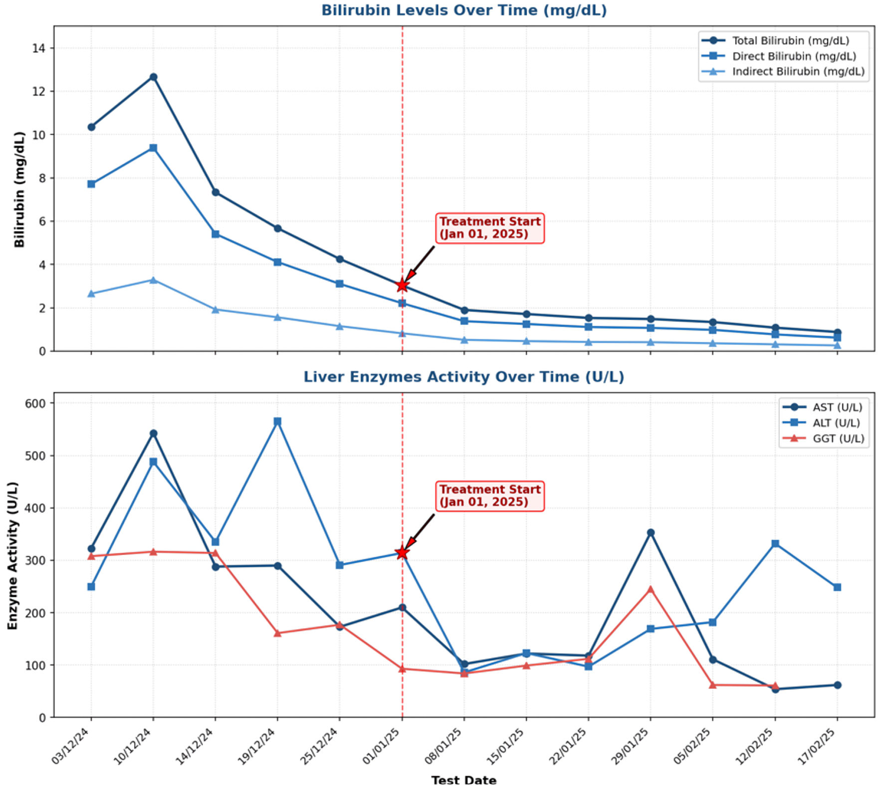 Click for large image | Figure 1. Liver biochemical profile with elevated aminotransferases and cholestatic enzymes. AST: aspartate aminotransferase; ALT: alanine aminotransferase; GGT: gamma-glutamyl transferase. |
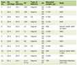 Click to view | Table 1. Approach to Prolonged Neonatal Cholestasis in a 2-Month-Old Infant |
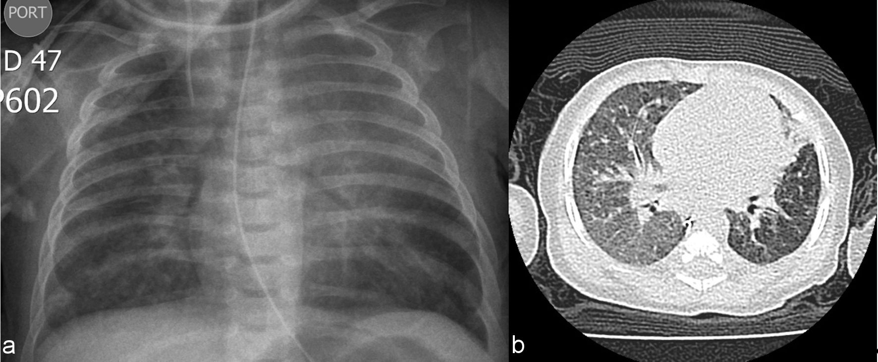 Click for large image | Figure 2. Baseline chest X-ray and CT. (a) Bilateral thickening of the peribronchovascular parahilar interstitium with consolidation in the upper lobe and lingula. (b) Chest computed tomography with multilobar involvement characterized by bilateral ground-glass opacities, peripheral micronodules, and alveolar filling regions. CT: computed tomography. |
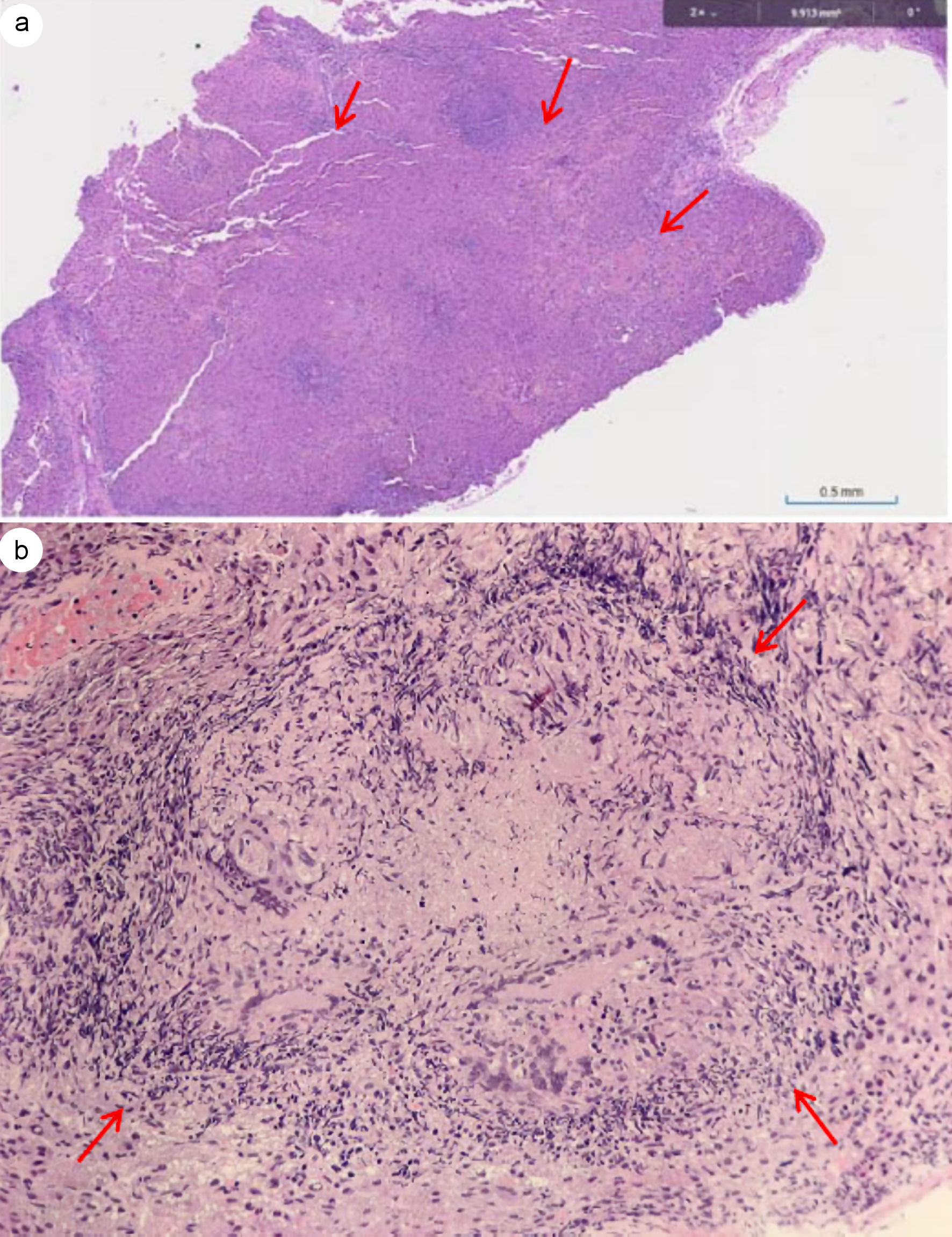 Click for large image | Figure 3. Liver biopsy (a) Multiple granulomas in the liver parenchyma (arrows, H&E, × 10). (b) Granuloma with peripheral halo of lymphocytes, central necrosis, and presence of multinucleated giant cells (arrows, H&E, × 40). H&E: hematoxylin and eosin stain. |
| Discussion | ▴Top |
Granulomatous hepatitis in the pediatric population is a rare entity with a heterogeneous and frequently nonspecific clinical presentation, typically characterized by prolonged fever, hepatomegaly, and abnormalities in liver function tests. Its reported prevalence has declined over time, from approximately 4% in earlier cohorts to less than 0.6% in more recent series [6]. Within this framework, miliary tuberculosis in infants constitutes an uncommon yet highly severe form of disease, driven by extensive hematogenous dissemination of M. tuberculosis. Although it represents only 1–7% of pediatric tuberculosis cases, it carries a disproportionately high mortality risk and is often characterized by a subacute course with failure to thrive, hepatosplenomegaly, and progressive respiratory compromise [7–9].
This case is notable for its initial presentation as prolonged neonatal cholestasis, which obscured the underlying pulmonary involvement until the onset of acute respiratory failure. Although the lungs constitute the primary site of miliary dissemination—supported by the characteristic CT findings of diffuse micronodules and ground-glass opacities reflecting alveolar and interstitial inflammation due to hematogenous spread—our patient remained previously healthy, without evidence of immunodeficiency or typical features such as prolonged fever or constitutional symptoms [10, 11]. Remarkably, cholestasis, defined by a significant elevation of conjugated bilirubin (> 1.0 mg/dL) [12], represented the sole manifestation of hepatic involvement. During the initial evaluation, the most common infectious etiologies for this age group were systematically excluded, and there was no history of perinatal exposure or maternal active tuberculosis [10]. In this context, histopathological findings on liver biopsy were pivotal in raising suspicion for tuberculosis, which was subsequently supported by microbiological confirmation and the identification of active tuberculosis in the patient’s father as the likely source of exposure.
Hepatic granulomas are found in 2–15% of liver biopsies, with more than 60% secondary to systemic processes; in developing countries, infectious etiologies have a higher incidence. However, tuberculous etiology in this context is uncommon, and most cases reported in the literature have been identified in patients with human immunodeficiency virus (HIV) or other states of immunosuppression [11, 13]. Tuberculous granulomas are characterized by central caseous necrosis and prominent peripheral inflammatory infiltrate, distinguishing them from sarcoid, suppurative, foreign body, and xanthomatous granulomas. The immune response involves the activation of macrophages and helper T lymphocytes toward Th1, Th2, Th17, or T-regulatory subpopulations [11].
The treatment should follow the recommendations established by the World Health Organization, ensuring full compliance with the anti-tuberculosis regimen [14]. Patients with hepatitis secondary to tuberculosis have shown an excellent therapeutic response, with resolution of liver lesions and significant clinical improvement.
Conclusions
Disseminated tuberculosis should be considered in infants with unexplained prolonged cholestasis or granulomatous hepatitis, particularly when respiratory deterioration, characteristic pulmonary imaging findings, or a potential household tuberculosis exposure is present. In this setting, integrating hepatic histopathology with thoracic imaging and microbiological testing may facilitate early recognition of miliary tuberculosis. Prompt initiation of appropriate antituberculosis therapy is essential to prevent progression of disseminated disease and improve clinical outcomes.
Acknowledgments
We thank the genetics and rheumatology departments for their diagnostic support.
Financial Disclosure
The authors declare that they have not received any funding for the preparation of this manuscript.
Conflict of Interest
The authors declare that they have no conflict of interest.
Informed Consent
Written informed consent was obtained from the patient’s legal guardian for the publication of this case report, in accordance with the institutional guidelines of Fundacion Santa Fe de Bogota. Patient anonymity and confidentiality have been strictly maintained.
Author Contributions
CZP and JFVC conceived and designed the case report. CZ, MJRB, JCM, MAS, KG, and SDRA contributed to patient management, investigation, and data collection. RLP and KG contributed to histopathological evaluation and interpretation. JFVC and OPP provided clinical supervision and contributed to the interpretation of the findings. MJRB and CZ performed the literature review and drafted the manuscript. All authors contributed to the critical revision of the manuscript, approved the final version, and agreed to be accountable for all aspects of the work.
Data Availability
Any inquiries regarding supporting data availability of this study should be directed to the corresponding author.
| References | ▴Top |
- Moore BK, Graham SM, Nandakumar S, Doyle J, Maloney SA. Pediatric tuberculosis: a review of evidence-based best practices for clinicians and health care providers. Pathogens. 2024;13(6):467.
doi pubmed - Evans RP, Mourad MM, Dvorkin L, Bramhall SR. Hepatic and Intra-abdominal Tuberculosis: 2016 Update. Curr Infect Dis Rep. 2016;18(12):45.
doi pubmed - Carrol ED, Clark JE, Cant AJ. Non-pulmonary tuberculosis. Paediatr Respir Rev. 2001;2(2):113-119.
doi pubmed - Kreft Vega J, Guarda Barros ME, Cox Fuenzalida PP. Diagnostico de tuberculosis en pediatria. Neumol Pediatrica. 2024;19(3):97-102.
doi - Basile FW, Nabeta P, Ruhwald M, Song R. Pediatric tuberculosis diagnostics: present and future. J Pediatric Infect Dis Soc. 2022;11(Supplement_3):S85-S93.
doi pubmed - Shaheen M, Lei GS, Relich RF, Jarasvaraparn C, Tolliver KM, Molleston JP, Gonzalez IA. Granulomas in pediatric liver biopsies: single center experience. Pediatr Dev Pathol. 2024;27(3):218-227.
doi pubmed - Jaganath D, Beaudry J, Salazar-Austin N. Tuberculosis in children. Infect Dis Clin North Am. 2022;36(1):49-71.
doi pubmed - Marais BJ, Gie RP, Schaaf HS, Hesseling AC, Obihara CC, Starke JJ, Enarson DA, et al. The natural history of childhood intra-thoracic tuberculosis: a critical review of literature from the pre-chemotherapy era. Int J Tuberc Lung Dis. 2004;8(4):392-402.
pubmed - Kumar NS, et al. Miliary tuberculosis: an atypical presentation. Int J Contemp Pediatr. 2021;8(6):1111.
doi - Chen HL, Taylor SA, Lee WS, Ciocca M, El-Guindi MA, Yachha SK, Fawaz R, et al. Diagnostic approaches for infants with cholestatic liver diseases: Position paper and perspectives of the Federation of International Societies of Pediatric Gastroenterology, Hepatology, and Nutrition. J Pediatr Gastroenterol Nutr. 2025;81(5):1360-1377.
doi pubmed - Ortiz-Brizuela E, Azamar-Llamas D, Mora JD, Guerrero-Castillo JI, Martinez-Benitez B, Sifuentes-Osornio J. Enfermedad granulomatosa hepatica. Gac Med Mex. 2019;155(3):266-275.
doi pubmed - Fawaz R, Baumann U, Ekong U, Fischler B, Hadzic N, Mack CL, McLin VA, et al. Guideline for the evaluation of cholestatic jaundice in infants: joint recommendations of the North American Society for Pediatric Gastroenterology, Hepatology, and Nutrition and the European Society for Pediatric Gastroenterology, Hepatology, and Nutrition. J Pediatr Gastroenterol Nutr. 2017;64(1):154-168.
doi pubmed - Hickey AJ, Gounder L, Moosa MY, Drain PK. A systematic review of hepatic tuberculosis with considerations in human immunodeficiency virus co-infection. BMC Infect Dis. 2015;15:209.
doi pubmed - WHO consolidated guidelines on tuberculosis: Module 5: Management of tuberculosis in children and adolescents. in WHO Guidelines Approved by the Guidelines Review Committee. Geneva: World Health Organization, 2022. Accessed: October 20, 2025. [Online]. Available: http://www.ncbi.nlm.nih.gov/books/NBK579387/.
This article is distributed under the terms of the Creative Commons Attribution 4.0 International License (CC BY 4.0), which permits unrestricted use, distribution, and reproduction in any medium, including commercial use, provided the original work is properly cited.
Journal of Medical Cases is published by Elmer Press Inc.