| Journal of Medical Cases, ISSN 1923-4155 print, 1923-4163 online, Open Access |
| Article copyright, the authors; Journal compilation copyright, J Med Cases and Elmer Press Inc |
| Journal website https://jmc.elmerpub.com |
Case Report
Volume 18, 2027, e5390

Incidental Discovery of Dual Intrauterine Devices With Bladder Perforation Involving One Device
Aminah Al-Khuder
Department of Obstetrics and Gynecology, Kuwait Institute for Medical Specialization (KIMS), Kuwait
Manuscript submitted June 26, 2026, accepted November 6, 2026, published online November 20, 2026
Short title: Incidental Dual IUDs With Bladder Perforation
doi: https://doi.org/10.14740/jmc5390
| Abstract | ▴Top |
Intrauterine contraceptive devices (IUDs) are highly effective and widely used long-acting reversible contraceptives. Uterine perforation is an uncommon complication, reported in approximately 1–2 per 1,000 insertions, and may result in extrauterine migration to adjacent organs. Bladder involvement is sporadic and may remain clinically silent, leading to delayed or incidental diagnosis. A 40-year-old multiparous woman presented with intermittent painless hematuria several years after IUD insertion. Following postpartum insertion of a copper IUD, the device was not visualized on repeated ultrasonography and was assumed expelled after an unremarkable dilation and curettage. A second hormonal IUD was subsequently inserted. Years later, imaging revealed the presence of two IUDs: one had perforated the uterine wall and embedded into the urinary bladder wall, while the second was partially perforating the uterus. Surgical removal via laparotomy with bladder repair was successfully performed, along with bilateral tubal ligation. This case highlights the diagnostic challenges associated with presumed IUD expulsion and delayed recognition of X-ray time migration. It emphasizes the importance of appropriate imaging to confirm the location of missing IUD before reinsertion and the need to consider device migration in patients presenting with unexplained urinary symptoms.
Keywords: Intrauterine contraceptive device; Urinary bladder injuries; Device migration; Uterine perforation; Incidental findings
| Introduction | ▴Top |
Intrauterine contraceptive devices (IUDs) are among the most effective long-term reversible contraceptive methods, with high continuation rates and an excellent overall safety profile. According to guidance from the American College of Obstetricians and Gynecologists (ACOG) and the Royal College of Obstetricians and Gynaecologists (RCOG), uterine perforation is an uncommon but recognized complication of IUD insertion, with reported incidence of approximately 0.1–0.3% [1–4].
Uterine perforations may occur during insertion and remain clinically unregonized for prolonged periods [3, 4].
Extrauterine migration can involve adjacent pelvic organs, including the urinary bladder, although such involvement is uncommon. Postpartum insertion and breastfeeding have been associated with an increased risk of uterine perforation [3, 4].
When IUD strings are not visualized, possible causes include string retraction, expulsion, and uterine perforation.
Appropriate imaging is essential to establish the location of the device and exclude extrauterine migration before assuming expulsion or considering reinsertion [5, 6]. Although intravesical IUD migration and the presence of multiple IUDs have been previously reported, the present case is striking for the coexistence of two devices involving different anatomical locations, including uterine perforation and bladder involvement. The patient presented with intermittent painless hematuria several years after presumed expulsion of the initial device and subsequent insertion of a second IUD. This case highlights the potential consequences of unrecognized device migration and emphasizes the importance of confirming the location of a missing IUD before reinsertion, as well as considering extrauterine migration in patients presenting with unexplained urinary symptoms.
| Case Report | ▴Top |
Investigations
A 40-year-old woman, para 4, all delivered vaginally, with no known medical illnesses, presented for evaluation of abnormal pelvic imaging findings. Her surgical history included abdominoplasty, perineorrhaphy, and laparoscopic cholecystectomy. She reported regular menstrual cycles.
Following her last delivery, a copper IUD was inserted at a private clinic. Over the following year, she experienced intermittent postcoital spotting. Pelvic ultrasonography reportedly failed to visualize the IUD within the uterine cavity, and a repeat detailed ultrasound similarly did not demonstrate the device. She subsequently underwent dilation and curettage (D&C), during which the IUD was not identified, and the procedure was reported as unremarkable. No further imaging was performed, and the IUD was presumed absent.
Approximately 1 year later, a second (hormonal) IUD was inserted. Thereafter, she developed irregular menstrual cycles characterized by infrequent but heavy bleeding, for which she did not seek medical attention. She reported it was tolerable.
Several years later, she presented with intermittent painless hematuria without associated urinary symptoms. Initial investigations were unremarkable. Subsequent pelvic X-ray revealed the presence of two IUDs, one of which had perforated the uterine wall with migration into the urinary bladder, while the second was visualized perforating through the uterus. She was referred to our center for further management.
Diagnosis
Office cystoscopy preformed on February 19, 2025 demonstrated a hormonal IUD perforating the dome of the bladder with calcified intravesical components as shown in Figure 1.
 Click for large image | Figure 1. Cystoscopic view demonstrating a calcified intrauterine contraceptive device perforating the bladder wall. |
Subsequent cystogram and computed tomography (CT) urography confirmed migration of IUD anterior to the uterine fundus with perforation into the anterosuperior bladder wall, as well as second low-lying intrauterine device within the cervical canal. These findings confirmed dual IUD with bladder perforation.
Pre-operative urine analysis demonstrated significant hematuria (> 50 red blood cells (RBCs) per high-power filed) with no evidence of urinary tract infection (UTI). These findings correlated with the patient’s painless hematuria and the cystoscopic evidence of bladder perforation.
Treatment
The patient underwent surgical removal of both IUDs by laparotomy on March 20, 2025. The IUD (hormonal) within the uterus was first removed transvaginally, followed by laparotomy, which revealed a calcified extrauterine IUD perforating the urinary bladder as shown in Figures 2 and 3. The migrated device was excised in its entirety, and the bladder defect was repaired in two layers, with a negative water seal test. Following counseling regarding contraceptive options, the patient expressed a desire for permanent contraception as she considered her family complete. Given her traumatic experience with IUD use, intrauterine contraception was no longer acceptable. She declined oral contraceptive methods. Therefore, she opted for bilateral tubal ligation, which was performed alongside. This case highlights the importance of individualized contraceptive counseling following device-related complications. Clinicians should ensure that patients receive balanced information regarding all appropriate contraceptive options, while incorporating the patient’s reproductive goals, previous experiences, and informed preferences through a shared decision-making process.
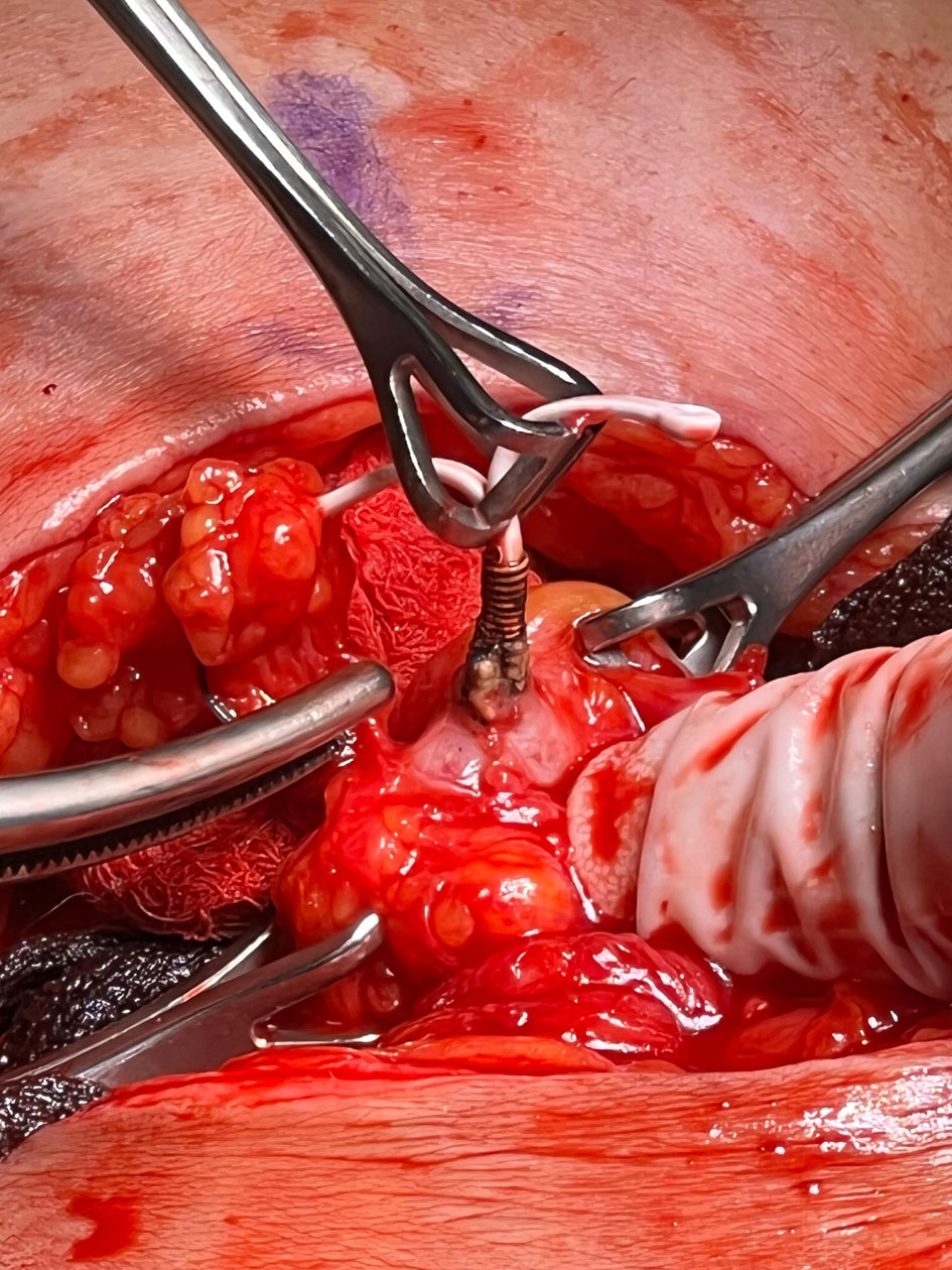 Click for large image | Figure 2. Intraoperative image demonstrating the removal of a second intrauterine contraceptive device via laparotomy. |
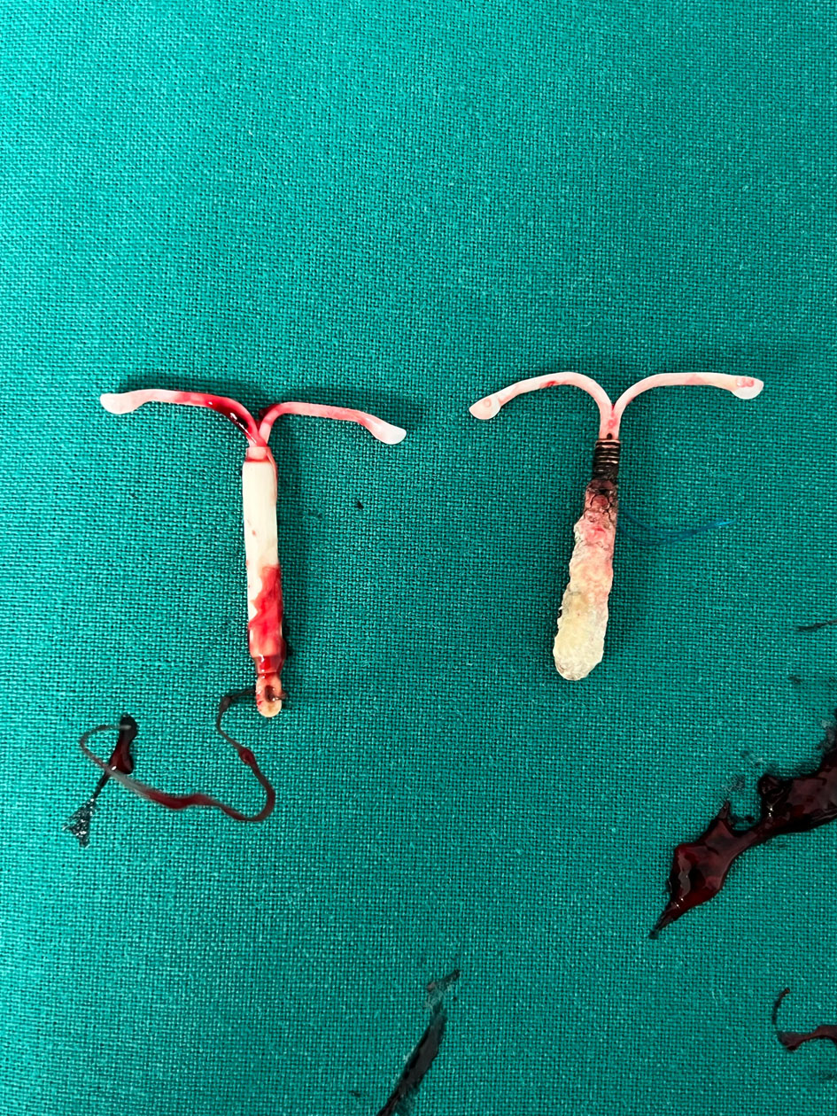 Click for large image | Figure 3. Retrieved intrauterine contraceptive devices displayed side by side, including a hormonal device and a copper device. |
Follow-up and outcomes
The postoperative course was uneventful. The patient was managed with antibiotics, analgesia, thromboprophylaxis, and bladder catheterization. Thromboprophylaxis was administered in accordance with institutional postoperative protocols during inpatient recovery, mainly in view of reduced postoperative mobility. She was scheduled for Foley’s catheter removal after 2 weeks and outpatient follow-up thereafter. Postoperative labs showed transient leukocytosis with neutrophilia, consistent with a postoperative inflammatory response:
- Hemoglobin and renal function remained stable;
- Urine culture showed no growth, with no evidence of UTI;
- Foley catheter removed after 2 weeks;
- At follow-up, the patient was clinically well and asymptomatic, with complete resolution of hematuria.
Patient perspective
The patient expressed sadness and disappointment upon discovering the presence of two IUDs and concerns regarding her previous medical evaluation. Following counseling about the available surgical options, she participated in shared decision-making and agreed to an open surgical approach for IUD removal, bilateral tubal ligation, and bladder repair. She expressed satisfaction with the overall management and treatment received.
| Discussion | ▴Top |
IUDs are among the most effective long-acting reversible contraceptive methods, with an excellent safety profile. Although uterine perforation and intravesical IUD migration are recognized complications, the present case shows a distinct clinical course involving two IUDs in different anatomical locations, delayed recognition of extrauterine migration, and subsequent presentation with intermittent painless hematuria. Its clinical significance lies primarily in the diagnostic challenges associated with presumed IUD expulsion and subsequent reinsertion without confirmation of the original device’s location. The case illustrates the potential consequences of presumed IUD expulsion without radiological confirmation, followed by insertion of a second device, and emphasizes the diagnostic challenges associated with delayed recognition of extrauterine migration.
The clinical significance of this case lies in the importance of appropriate investigation of a missing IUD before subsequent device insertion, as well as maintaining clinical suspicion for device migration in patients presenting with unexplained urinary symptoms given the history. Uterine perforations are uncommon, with a reported incidence of approximately 1.3 per 1,000 insertions [3].
It is believed to occur most commonly at the time of IUD insertion and is asymptomatic. In cases of symptomatic lower abdominal pain, non-visible threads and changes in bleeding may indicate uterine perforation.
Postpartum insertion and breastfeeding have been associated with an increased risk of uterine perforation [3, 7]. As the initial IUD in our patient was inserted following childbirth, postpartum uterine changes may have been a contributing factor; however, the timing and mechanism of perforation could not be established retrospectively.
This case highlights important diagnostic challenges associated with missing IUDs. Transvaginal ultrasonography is commonly used as the first-line investigation; however, it may fail to detect extrauterine devices. In this patient, repeated ultrasound examinations did not visualize the IUD within the uterine cavity, suggesting the device had been expelled. Especially, D&C was performed due to a mild endometrial thickness noticed by ultrasound and to provide direct visualization to confirm the availability of previously inserted IUDs, during which the device was not identified, and the procedure was reported as unremarkable. As shown in this case, D&C does not exclude extrauterine migration, and relying on it alone may delay diagnosis [5].
Current recommendations emphasize that when IUD strings are not visualized and the device is not identified within the uterus, we need to exclude pregnancy (pregnancy test), consider emergency contraception, and provide alternative contraception. An ultrasound should be arranged. If it is not in situ, a pelvic/abdomen radiography should be performed to confirm the presence or absence of the device prior to assuming expulsion or proceeding with reinsertion [2, 8].
Failure to perform confirmatory imaging in this case led to reinsertion of a second IUD while the first device remained extrauterine, ultimately resulting in dual IUDs. In line with Faculty of Sexual & Reproductive Healthcare guidance, when uterine perforation is known or suspected, reinsertion of IUD should be delayed for 6 weeks [2].
Delayed recognition of IUD migration has been documented in previous reports, with patients presenting years after insertion with persistent or recurrent urinary symptoms. One study described a case of intravesical migration diagnosed 15 years after insertion, following recurrent urinary tract symptoms and hematuria [9]. Prolonged asymptomatic IUD migration has also been described by Jeje et al, who reported an IUD discovered 20 years after insertion during investigation of hematuria [10]. Also, another similar case documented onset of urinary symptoms 13 years after device insertion, with exercise-induced hematuria as the predominant symptom [11]. Similarly, a previously reported case involving two IUDs showed how presumed expulsion of the initial device, followed by reinsertion, led to delayed identification of an extrauterine IUD involving the bladder [12].
Furthermore, a literature review of 115 cases of urinary tract IUD migration identified dysuria (33%), hematuria (26%), and UTI (16%) among reported clinical manifestations [13]. These findings parallel the present case, in which delayed recognition of IUD migration was followed by intermittent painless hematuria. Collectively, these reports emphasize that persistent or unexplained urinary symptoms in patients with a history of IUD insertion should prompt consideration of extrauterine migration. Clinical presentation of migrated IUDs is variable. Many patients remain asymptomatic for prolonged periods, while others may present with lower urinary tract symptoms, recurrent UTIs, bladder stone formation, or hematuria [6]. In this case, the patient presented several years later with intermittent painless hematuria, which was ultimately attributable to bladder wall perforation by the migrated device. The absence of infective features and negative urine cultures further supported a mechanical etiology.
Management of migrated IUDs depends on anatomical location of the device, degree of organ involvement, and associated complications. Although minimally invasive approaches may be appropriate in selected cases, open surgery was performed in our patient, allowing removal of both devices and repair of the bladder defect. The choice of surgical approach could be considered in relation to the operative findings and the anticipated need for organ repair.
This case also underscores the importance of contraceptive counseling and patient-centered decision-making. Following her adverse and distressing experience with IUD use, the patient declined further intrauterine or oral contraceptive methods and requested permanent contraception, having completed her family. After removal of both devices and bladder repair, bilateral tubal ligation using clips was performed for permanent contraception, according to the patient’s preference and the operating surgeon’s decision; bilateral salpingectomy was not performed, and a specific rationale for choosing tubal ligation over salpingectomy was not documented.
The most common location is the peritoneal cavity, although migration to adjacent organs such as the urinary bladder has been reported. The management strategy recommended by WHO is surgical removal by laparoscopic intervention [14]. Laparotomy may be indicated in case of sepsis, bowel perforation, or inability to remove the IUD by laparoscope.
Conclusion
This case highlights an exceptionally rare presentation of dual IUDs, one of which migrated extrauterine and perforated the urinary bladder, presenting years after insertion. It underscores the importance of confirming IUD location with appropriate imaging when strings are not visualized, as ultrasonography alone may be insufficient, and D&C does not exclude extrauterine migration. Early recognition and accurate localization are essential to prevent delayed complications. In addition, this case emphasizes the role of patient-centered contraceptive counseling, as prior adverse experiences may significantly influence future contraceptive choices.
Acknowledgments
None to declare.
Financial Disclosure
The author has no funding to declare.
Conflict of Interest
The author declares no conflict of interest.
Informed Consent
Written informed consent was obtained from the patient for publication of this case report and accompanying images.
Author Contributions
The author was responsible for the conception, design, data collection, analysis, and writing of the manuscript.
Data Availability
Data supporting the findings of this study are available from the corresponding author upon reasonable request.
| References | ▴Top |
- American College of O, Gynecologists' Committee on Gynecologic P, Long-Acting Reversible Contraceptive Expert Work G. Committee opinion No 672: clinical challenges of long-acting reversible contraceptive methods. Obstet Gynecol. 2016;128(3):e69-77.
doi pubmed - Faculty of sexual and reproductive healthcare. Intrauterine contraception. London: FSRH; 2023 (amended).
- Heinemann K, Reed S, Moehner S, Minh TD. Risk of uterine perforation with levonorgestrel-releasing and copper intrauterine devices in the European Active Surveillance Study on Intrauterine Devices. Contraception. 2015;91(4):274-279.
doi pubmed - Heinemann K, Curtis KM. Complications of intrauterine contraception. In: UpToDate. Waltham, MA: UpToDate Inc; 2024-2025.
- Boortz HE, Margolis DJ, Ragavendra N, Patel MK, Kadell BM. Migration of intrauterine devices: radiologic findings and implications for patient care. Radiographics. 2012;32(2):335-352.
doi pubmed - Rafique M. Intravesical migration of intrauterine device: case report and review of literature. Urol J. 2008;5(4):269-271.
- Kaislasuo J, Suhonen S, Gissler M, Lahteenmaki P, Heikinheimo O. Uterine perforation caused by intrauterine devices: clinical course and treatment. Hum Reprod. 2013;28(6):1546-1551.
doi pubmed - Curtis KM, Tepper NK, Jatlaoui TC, Berry-Bibee E, Horton LG, Zapata LB, Simmons KB, et al. U.S. medical eligibility criteria for contraceptive use, 2016. MMWR Recomm Rep. 2016;65(3):1-103.
doi pubmed - Salih MA, Tefera AT, Gebrehiwot FG, Mideksa AG, Halala NS, Gebreselassie KH. Gross hematuria caused by intravesical migration of a forgotten intrauterine device: a case report and literature review. Res Rep Urol. 2022;14:291-296.
doi pubmed - Jeje EA, Ojewola RW, Atoyebi OA. Intravesical migration of a failed and forgotten intrauterine contraceptive device after 20 years of insertion—a case report. Nig Q J Hosp Med. 2012;22(3):221-223.
pubmed - Mascarenhas MP, Tiraboschi RB, Paschoalin VP, Costa EA, Suzuki Bellucci CH, Bessa Junior J. Exercise-induced hematuria as the main manifestation of migration of intrauterine contraceptive device into the bladder. Case Rep Urol. 2012;2012:736426.
doi pubmed - Liu G, Li F, Ao M, Huang G. Intrauterine devices migrated into the bladder: two case reports and literature review. BMC Womens Health. 2021;21(1):301.
doi pubmed - Varlas VN, Meianu AI, Radoi AI, Balescu I, Bacalbasa N, Varlas RG. Intrauterine contraceptive device migrated in the urinary tract: case report and extensive literature review. J Clin Med. 2024;13(14).
doi pubmed - Mosley FR, Shahi N, Kurer MA. Elective surgical removal of migrated intrauterine contraceptive devices from within the peritoneal cavity: a comparison between open and laparoscopic removal. JSLS. 2012;16(2):236-241.
doi pubmed
This article is distributed under the terms of the Creative Commons Attribution 4.0 International License (CC BY 4.0), which permits unrestricted use, distribution, and reproduction in any medium, including commercial use, provided the original work is properly cited.
Journal of Medical Cases is published by Elmer Press Inc.