| Journal of Medical Cases, ISSN 1923-4155 print, 1923-4163 online, Open Access |
| Article copyright, the authors; Journal compilation copyright, J Med Cases and Elmer Press Inc |
| Journal website https://jmc.elmerpub.com |
Case Report
Volume 17, Number 10, October 2026, pages 585-589

Klebsiella pneumoniae Liver Abscess in a Filipino Patient With Previously Undiagnosed Diabetes: Successful Conservative Management
Luis Paz y Minoa, Manuel Calvopinaa, c, Julia Saaa, Pablo Echeverriab
aOne Health Research Group, Universidad de las Americas, Quito, Ecuador
bHSHS Saint Elizabeth’s Hospital, O’Fallon, IL, USA
cCorresponding Author: Manuel Calvopina, One Health Research Group, Universidad de las Americas, Quito, Ecuador
Manuscript submitted June 8, 2026, accepted July 31, 2026, published online September 4, 2026
Short title: K. pneumoniae Liver Abscess: Management
doi: https://doi.org/10.14740/jmc5377
| Abstract | ▴Top |
Klebsiella pneumoniae is a common pathogen of pyogenic liver abscess (PLA), particularly among individuals with diabetes mellitus and those of Asian ethnicity. A 32-year-old Filipino man with no prior medical history presented with fever, headache, nausea, and vomiting and was found to have new-onset diabetic ketoacidosis (DKA). Laboratory evaluation revealed leukocytosis, thrombocytopenia, elevated liver enzymes, hyperbilirubinemia and lactic acidosis. Abdominal computed tomography (CT) showed hepatosplenomegaly, variable hepatic abnormalities including < 2 cm hypodense lesions and a large ill-defined area of decreased density in the inferior right liver lobe. The patient was hospitalized for sepsis and DKA. Empiric piperacillin–tazobactam was administered for 24 h and subsequently de-escalated to ceftriaxone after blood cultures grew extended-spectrum β-lactamase–negative Klebsiella pneumoniae; ceftriaxone was continued for 4 weeks. Due to the number, size and inaccessible location of the liver lesions, percutaneous drainage was not feasible. After 16-week follow-up, the patient achieved complete clinical recovery and radiological resolution of hepatic lesions. This case highlights variable hepatic lesion presentations in Klebsiella liver abscess (KLA) with successful conservative management without drainage.
Keywords: Pyogenic liver abscess; Klebsiella liver abscess; Klebsiella pneumoniae
| Introduction | ▴Top |
Liver abscess is the most common type of visceral abscess [1]. While pyogenic liver abscess (PLA) predominates in developed countries, amebic/amoebic liver abscess (ALA) are prevalent in tropical endemic regions [2, 3]. Historically associated with high mortality, prompt diagnosis and management of PLA have reduced mortality rates from approximately 65% to 2–12% [1, 4]. Although PLA is frequently polymicrobial, causative organisms vary geographically. Streptococcus spp. and Escherichia coli seem to continue being the historical organisms in Western countries [5–7], whereas Klebsiella pneumoniae (K. pneumoniae) has been reported as the most common isolated pathogen in Asia, giving rise to Klebsiella liver abscess (KLA) [8, 9]. Nevertheless, KLA has increasingly been reported worldwide, raising concerns for its epidemiologic impact [6, 9, 10].
Most cases of KLA continue to occur in patients of Asian descent [10, 11]. Asian ethnicity may be a determinant for gastrointestinal colonization by some K. pneumoniae serotypes [10, 12]. Diabetes, however, remains one of the strongest predisposing factors for KLA [8, 13]. Some studies have described previously unrecognized diabetes diagnosed during hospitalization for PLA [14]. Nonetheless, indications for diabetes evaluation are still limited. Other risk factors associated with KLA include liver disease, gallbladder conditions, and malignancy [8, 15, 16].
Clinical and radiologic manifestations of PLA may be variable and occasionally mimic hepatic malignancy, posing diagnostic challenges [1]. Diagnosis relies primarily on imaging methods such as ultrasound or computed tomography (CT) with contrast-enhanced as the preferred imaging modality [17, 18]. KLA has more commonly been associated with the presentation of a solitary and relatively large abscess [16, 19]. Once PLA is highly suspected, aspirate cultures from the lesion should be obtained [15, 20–22]. However, some abscesses may not be amenable to percutaneous drainage, thereby limiting both diagnostic confirmation and the delivery of targeted therapy.
We report a case of KLA in a young Filipino patient with no known prior medical history who presented with diabetic ketoacidosis (DKA) and atypical hepatic lesions concerning for malignancy. The patient was successfully managed with antimicrobial therapy alone since percutaneous drainage was not technically feasible.
| Case Report | ▴Top |
A 32-year-old Filipino male with no known prior medical history presented to the hospital with a 2-day history of headache, nausea, vomiting, and fever. On admission, laboratory evaluation revealed metabolic acidosis with a pH of 7.31 (reference range: 7.35–7.45) and anion gap of 21.3 (8–12 mmol/L) plus marked hyperglycemia of 286 mg/dL (70–99 mg/dL), consistent with DKA. Moreover, glycosylated hemoglobin was elevated to 11.5% (< 5.7%), suggesting previously undiagnosed diabetes.
Given persistent fever along with headache, a lumbar puncture was warranted during admission, and its results were unremarkable. Laboratory studies showed leukocytosis 15.8 × 10⁹/L (4.0–11.0 × 10⁹/L) with bandemia, thrombocytopenia 92 × 109/L (150–400 × 109/L), elevated total bilirubin 1.7 mg/dL (0.1–1.2 mg/dL) and transaminases with aspartate aminotransferase (AST) of 171 U/L and alanine aminotransferase (ALT) of 124 U/L (AST 10–40 U/L; ALT 7–56 U/L), lactic acidosis 2.2 mmol/L (0.5–2.0 mmol/L) and normal urine analysis. Chest CT reported a small left pleural effusion. CT of the abdomen demonstrated hepatosplenomegaly and multiple hypodense lesions within the liver measuring less than 2 cm, and a large ill-defined area of decreased density in the inferior right liver lobe with slight/non-substantial enhancement changes, which raised concern for underlying malignancy (Fig. 1).
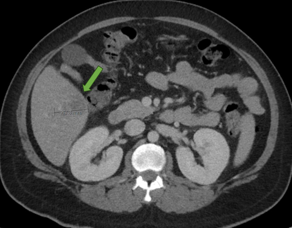 Click for large image | Figure 1. Contrast-enhanced axial abdominal CT image of the abdomen showing a 43.8-mm ill-defined hypoattenuating lesion in the inferior right hepatic lobe. The arrow identifies the larger hepatic lesion. CT: computed tomography. |
On hospital day 1, the patient was admitted to the intensive care unit for management of severe sepsis with shock and DKA. Empiric antibiotic therapy with piperacillin–tazobactam was initiated due to concern for possible liver infection noted on imaging. Within 24 h of admission, blood cultures grew K. pneumoniae (extended-spectrum beta-lactamase negative), prompting de-escalation of antimicrobial therapy to intravenous ceftriaxone. Extensive infectious workups, including stool cultures, Shiga toxin, Cryptosporidium antigen, parasite ova, amoebic serology, hepatitis B and C tests were performed, and all results were negative.
Radiology specialists argued that hepatic lesions were deemed not amenable for percutaneous drainage given their multiplicity, small size and location. At that time, no other symptoms were reported, and ocular examination was unremarkable. Therefore, the patient was managed conservatively with prolonged antimicrobial therapy and favorable clinical response. On hospital day 3, the DKA resolved, and he was transferred from the intensive care unit. Subsequently, he was discharged home on hospital day 8 with outpatient intravenous ceftriaxone. At week 3, repeat CT imaging showed minimal change in hepatic lesions. At week 4, he completed a total 4-week course of intravenous ceftriaxone. Finally, follow-up CT imaging obtained 16 weeks after admission as part of primary care monitoring demonstrated complete resolution of all liver lesions (Fig. 2). The patient remained asymptomatic with no clinical evidence of recurrence. Furthermore, basal insulin was prescribed for type 2 diabetes mellitus and adequate glycemic control was maintained with metformin alone 3 months after discharge. The patient provided verbal consent for academic publication of this case.
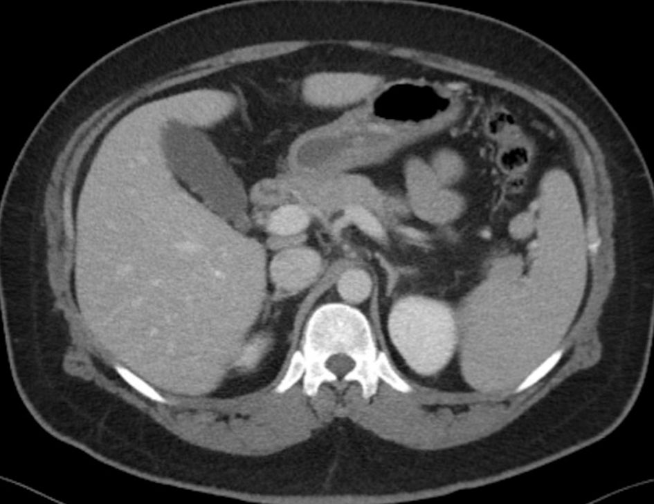 Click for large image | Figure 2. Follow-up contrast-enhanced axial CT image of the abdomen obtained 16 weeks after admission, showing complete radiological resolution with no residual hepatic lesions. CT: computed tomography. |
| Discussion | ▴Top |
The present case illustrates several remarkable features of PLA caused by K. pneumoniae. First of all, it highlights the emergence of K. pneumoniae as a pathogen causing PLA in the United States (USA). Secondly, it demonstrates the strong association between KLA and previously undiagnosed diabetes mellitus, including severe metabolic presentations such as DKA. Lastly, it shows that selected patients with multiple, small non-loculated abscesses not amenable to percutaneous drainage may achieve complete clinical and radiological resolution with antimicrobial therapy and careful monitoring. Our patient was originally from the Philippines, a region where KLA is well recognized, supporting previous observations that ethnicity and geographic background may influence gastrointestinal colonization with K. pneumoniae [10, 12].
Furthermore, this KLA case exemplifies the concomitant diagnosis of type 2 diabetes mellitus presenting as DKA. Diabetes is considered one of the strongest risk factors for KLA and has consistently been associated with increased susceptibility to invasive infection [8, 11, 13, 19]. Hyperglycemia may impair neutrophil chemotaxis, phagocytosis, and intracellular killing, facilitating dissemination of encapsulated organisms such as K. pneumoniae [8]. Reports have described patients in whom diabetes was first recognized during hospitalization for liver abscess [14], like our patient who had no previous diagnosis of diabetes. A glycated hemoglobin (HbA1c) of 11.5%, marked hyperglycemia of 286 mg/dL, and DKA led to a new diagnosis of type 2 diabetes mellitus. This previously unrecognized diabetes may have increased the patient’s susceptibility to K. pneumoniae [23]. Therefore, screening for diabetes should be considered in patients presenting with KLA.
KLA can occasionally mimic hepatic neoplasms, particularly when lesions appear infiltrative, irregular or when extensive necrosis is present on imaging studies [1, 24–26]. Liver malignancy suspicion may lead to delays in KLA recognition or unnecessary invasive procedures. Further evaluation using supportive advanced imaging could narrow the differential diagnosis, with features favoring hepatic abscess including a layered wall appearance with persistent rim enhancement, transient segmental enhancement, apparent diffusion coefficient (ADC) mapping. When the diagnosis remains uncertain, biopsy may provide definitive confirmation [1, 27, 28]. However, the hepatic lesions in this case showed only minimal enhancement, and ADC mapping—which is not routinely used or standardized for diagnosing PLA—was not obtained. In endemic regions and patients with compatible epidemiological exposures, ALA should also be included in the differential diagnosis [2, 3]. In our patient ALA diagnosis was excluded given the negative serology result. Risk factors for hepatic neoplasm were limited, and positive blood cultures for K. pneumoniae, together with clinical improvement after targeted antibacterial therapy, strongly supported the diagnosis of KLA.
Finally, prompt management is crucial, particularly given increasing rates of antibiotics resistance and higher risk of complications such as K. pneumoniae invasive liver abscess syndrome, cryptogenic KLA, and endogenous endophthalmitis [10, 11, 13, 19, 23]. In this case, cerebrospinal fluid analysis was normal, as well as ophthalmologic and chest examinations; hence no other metastatic foci of infection were identified. Nonetheless, clinicians should maintain a high index of suspicion for disseminated disease, particularly in diabetic patients [10, 11, 13, 19, 23]. Current management of PLA generally includes both antimicrobial therapy and abscess drainage, particularly for lesions that are large, multiloculated, or poorly responsive to antibiotics [1, 16, 18, 19]. Nevertheless, drainage is not always technically feasible, particularly in patients with multiple small lesions or lesions in difficult-to-access anatomical locations, as occurred in this case when the radiology team determined lesions not amenable for aspiration. Some reports suggest that selected patients with small abscesses may respond adequately to antibiotic therapy alone [1, 18]. While conservative antimicrobial management may be successful in selected patients, there is currently no universal guidance regarding lesion size thresholds (ranging from 3 to 5 cm of diameter) for drainage approaches [1, 18]. In addition, surgical intervention is generally reserved for cases in which percutaneous drainage fails to achieve adequate resolution [1, 18]. In this case, the patient had multiple lesions measuring less than 2 cm and one of 4 cm, all without loculation, and showed rapid clinical improvement after initiation of ceftriaxone. Complete radiological resolution after 16 weeks of antimicrobial therapy further supports the feasibility of non-interventional management in selected patients together with careful monitoring. Overall, KLA demands a multidisciplinary strategy encompassing clinical, radiological, microbiological, and even public health domains.
Conclusions
In conclusion, this case highlights the evolving epidemiology of KLA in Western countries and underscores the importance of considering K. pneumoniae liver abscess in patients of Asian origin presenting with sepsis and newly diagnosed diabetes mellitus. KLA may present with variable imaging findings, and patients with multiple, small, nonloculated non-drainable lesions could be managed successfully with antimicrobial therapy alone and careful monitoring. Early recognition, prompt first-line antimicrobial therapy, evaluation for underlying diabetes, and close radiological follow-up remain essential components of management.
Learning points
KLA continues spreading beyond Asia. Risk factors for KLA need to be assessed for differential diagnostic approaches, including Asian descent and diabetes. Atypical radiologic presentations pose diagnostic challenges, especially when mimicking malignancy. Abscesses may not be amenable to drainage when therapy relies on antibiotics and close monitoring.
Acknowledgments
None to declare.
Financial Disclosure
None to declare.
Conflict of Interest
None to declare.
Informed Consent
Verbal informed consent was obtained from the patient for publication of this totally anonymized case report. Despite multiple attempts, we have been unable to obtain written informed consent due to a failure to reach the patient.
Author Contributions
Direct patient care, review and writing of drafts (Pablo Echeverria); preparation and writing of manuscript (Luis Paz y Mino); review and writing of draft/manuscript (Manual Calvopina); conceptualization and investigation (Julia Saa).
Data Availability
The data supporting the findings of this study are available from the corresponding author upon reasonable request.
AI Use Declaration
During the preparation of this work, the authors used ChatGPT to edit spelling and punctuation. After using this tool, the authors reviewed and edited the content.
Abbreviations
PLA: pyogenic liver abscess; KLA: Klebsiella liver abscess; ALA: amebic/amoebic liver abscess; DKA: diabetic ketoacidosis; CT: computed tomography; ADC: apparent diffusion coefficient; K. pneumoniae: Klebsiella pneumoniae
| References | ▴Top |
- Bachler P, Baladron MJ, Menias C, Beddings I, Loch R, Zalaquett E, Vargas M, et al. Multimodality imaging of liver infections: differential diagnosis and potential pitfalls. Radiographics. 2016;36(4):1001-1023.
doi pubmed - Chavez-Tapia NC, Hernandez-Calleros J, Tellez-Avila FI, Torre A, Uribe M. Image-guided percutaneous procedure plus metronidazole versus metronidazole alone for uncomplicated amoebic liver abscess. Cochrane Database Syst Rev. 2009;2009(1):CD004886.
doi pubmed - Honecker B, Hansen CS, Lotter H. Sex in immune cells and parasitic diseases - a complex relationship. Immunol Rev. 2026;337(1):e70097.
doi pubmed - Meddings L, Myers RP, Hubbard J, Shaheen AA, Laupland KB, Dixon E, Coffin C, et al. A population-based study of pyogenic liver abscesses in the United States: incidence, mortality, and temporal trends. Am J Gastroenterol. 2010;105(1):117-124.
doi pubmed - Cerwenka H. Pyogenic liver abscess: differences in etiology and treatment in Southeast Asia and Central Europe. World J Gastroenterol. 2010;16(20):2458-2462.
doi pubmed - Mukthinuthalapati V, Attar BM, Parra-Rodriguez L, Cabrera NL, Araujo T, Gandhi S. Risk factors, management, and outcomes of pyogenic liver abscess in a US safety net hospital. Dig Dis Sci. 2020;65(5):1529-1538.
doi pubmed - Kaplan GG, Gregson DB, Laupland KB. Population-based study of the epidemiology of and the risk factors for pyogenic liver abscess. Clin Gastroenterol Hepatol. 2004;2(11):1032-1038.
doi pubmed - Hasan GM, Mohammad T, Zaidi S, Shamsi A, Sohal SS, Hassan MI. <p>Klebsiella pneumoniae and pyogenic liver abscess: Emerging clinical threats, virulence mechanisms and therapeutic strategies (Review)</p>. Mol Med Rep. 2026;33(3):90.
doi pubmed - Yin D, Ji C, Zhang S, Wang J, Lu Z, Song X, Jiang H, et al. Clinical characteristics and management of 1572 patients with pyogenic liver abscess: A 12-year retrospective study. Liver Int. 2021;41(4):810-818.
doi pubmed - Siu LK, Yeh KM, Lin JC, Fung CP, Chang FY. Klebsiella pneumoniae liver abscess: a new invasive syndrome. Lancet Infect Dis. 2012;12(11):881-887.
doi pubmed - Cardenas-Alvarez J, Balayla G, Triana A, Diaz Lankenau R, Franco-Paredes C, Henao-Martinez AF, Motoa G. Clinical spectrum and outcomes of cryptogenic Klebsiella pneumoniae liver abscess in the Americas: a scoping review. Pathogens. 2023;12(5).
doi pubmed - Lin YT, Siu LK, Lin JC, Chen TL, Tseng CP, Yeh KM, Chang FY, et al. Seroepidemiology of Klebsiella pneumoniae colonizing the intestinal tract of healthy Chinese and overseas Chinese adults in Asian countries. BMC Microbiol. 2012;12:13.
doi pubmed - Zhang L, Chen J, Qu Y, Cao X, Cui J, Li J, Yu A. Development and validation of a predictive model for invasive syndrome in patients with Klebsiella pneumoniae liver abscess. Front Med (Lausanne). 2025;12:1663407.
doi pubmed - Lin YT, Wang FD, Wu PF, Fung CP. Klebsiella pneumoniae liver abscess in diabetic patients: association of glycemic control with the clinical characteristics. BMC Infect Dis. 2013;13:56.
doi pubmed - Li S, Yu S, Peng M, Qin J, Xu C, Qian J, He M, et al. Clinical features and development of Sepsis in Klebsiella pneumoniae infected liver abscess patients: a retrospective analysis of 135 cases. BMC Infect Dis. 2021;21(1):597.
doi pubmed - Liu J, Liu Y, Li C, Peng W, Jiang C, Peng S, Fu L. Characteristics of Klebsiella pneumoniae pyogenic liver abscess from 2010-2021 in a tertiary teaching hospital of South China. J Glob Antimicrob Resist. 2024;36:210-216.
doi pubmed - Webb GJ, Chapman TP, Cadman PJ, Gorard DA. Pyogenic liver abscess. Frontline Gastroenterol. 2014;5(1):60-67.
doi pubmed - Mavilia MG, Molina M, Wu GY. The evolving nature of hepatic abscess: a review. J Clin Transl Hepatol. 2016;4(2):158-168.
doi pubmed - Chan KS, Chia CTW, Shelat VG. Demographics, radiological findings, and clinical outcomes of klebsiella pneumonia vs. non-klebsiella pneumoniae pyogenic liver abscess: a systematic review and meta-analysis with trial sequential analysis. Pathogens. 2022;11(9):976.
doi pubmed - Wang JH, Liu YC, Lee SS, Yen MY, Chen YS, Wang JH, Wann SR, et al. Primary liver abscess due to Klebsiella pneumoniae in Taiwan. Clin Infect Dis. 1998;26(6):1434-1438.
doi pubmed - Molton JS, Chan M, Kalimuddin S, Oon J, Young BE, Low JG, Salada BMA, et al. Oral vs intravenous antibiotics for patients with klebsiella pneumoniae liver abscess: a randomized, controlled noninferiority study. Clin Infect Dis. 2020;71(4):952-959.
doi pubmed - Miller JM, Binnicker MJ, Campbell S, Carroll KC, Chapin KC, Gonzalez MD, Harrington A, et al. Guide to utilization of the microbiology laboratory for diagnosis of infectious diseases: 2024 update by the Infectious Diseases Society of America (IDSA) and the American Society for Microbiology (ASM). Clin Infect Dis. 2024.
doi pubmed - Chen KY, Chan HC, Chan CM. How does hypervirulent Klebsiella pneumoniae influence the risk and visual outcomes of endogenous endophthalmitis in Klebsiella pneumoniae pyogenic liver abscess? A systematic review and meta analysis. J Infect Public Health. 2026;19(3):103090.
doi pubmed - Wang CH, Sun CK, Jiang JS, Tsai MH. Tumor-like liver abscess mimicking malignancy with lung metastases in a patient with acute renal failure: a case report. Medicine (Baltimore). 2016;95(11):e3145.
doi pubmed - Xu J, Wu D, Wang P, Zhou H. Klebsiella pneumoniae liver abscess mimicking cholangiocarcinoma: A case report. Asian J Surg. 2022;45(12):2750-2751.
doi pubmed - Ozgul E. Multiple pyogenic liver abscesses mimicking metastatic disease on computed tomography. Cureus. 2020;12(2):e7050.
doi pubmed - Shah S, Shukla A, Paunipagar B. Radiological features of hepatocellular carcinoma. J Clin Exp Hepatol. 2014;4(Suppl 3):S63-66.
doi pubmed - Chernyak V, Fowler KJ, Kamaya A, Kielar AZ, Elsayes KM, Bashir MR, Kono Y, et al. Liver imaging reporting and data system (LI-RADS) version 2018: imaging of hepatocellular carcinoma in at-risk patients. Radiology. 2018;289(3):816-830.
doi pubmed
This article is distributed under the terms of the Creative Commons Attribution 4.0 International License (CC BY 4.0), which permits unrestricted use, distribution, and reproduction in any medium, including commercial use, provided the original work is properly cited.
Journal of Medical Cases is published by Elmer Press Inc.