| Journal of Medical Cases, ISSN 1923-4155 print, 1923-4163 online, Open Access |
| Article copyright, the authors; Journal compilation copyright, J Med Cases and Elmer Press Inc |
| Journal website https://jmc.elmerpub.com |
Case Report
Volume 000, Number 000, May 2026, pages 000-000

A Case of Peripartum Cardiogenic Shock Resulting From Reverse Takotsubo Cardiomyopathy
Nazish Tarara, e, Hannah Walshb, Katherine Sadaniantza, Shantel Brissettc, Anna R. Wheland, Lara Kovellc
aDepartment of Internal Medicine, UMass Chan Medical School, Worcester MA, USA
bUMass Chan Medical School, Worcester MA, USA
cDepartment of Cardiovascular Medicine, UMass Chan Medical School, Worcester MA, USA
dDepartment of Obstetrics and Gynecology, UMass Chan Medical School, Worcester MA, USA
eCorresponding Author: Nazish Tarar, Department of Internal Medicine, UMass Chan Medical School, Worcester MA 01605, USA
Manuscript submitted March 5, 2026, accepted May 5, 2026, published online May 27, 2026
Short title: Complex Case of Reverse Stress Cardiomyopathy
doi: https://doi.org/10.14740/jmc5324
| Abstract | ▴Top |
Takotsubo cardiomyopathy (TCM) is characterized by transient left ventricular (LV) dysfunction that classically presents as apical ballooning with basal hyperkinesis. A recognized variant, reverse TCM, conversely is characterized by basal and mid-ventricular hypokinesis with apical sparing, often triggered by a catecholamine surge. We present a case of a woman who underwent an emergency cesarean section complicated by the development of cardiogenic shock due to TCM. She had rapid clinical improvement with restoration of systolic function and was ultimately treated with guideline-directed medical therapy with a multidisciplinary team to ensure safety while lactating. In this case, the acute onset of LV systolic dysfunction in the setting of physical stress, wall motion abnormalities in a non-ischemic distribution, and rapid clinical recovery were factors that favored TCM as a diagnosis. The diagnosis of pregnancy-associated TCM is challenging, yet differentiation from other cardiomyopathies is important to guide acute management, as well as prognosis and counseling for subsequent pregnancies.
Keywords: Takotsubo cardiomyopathy; Stress cardiomyopathy; Peripartum cardiogenic shock; Pregnancy
| Introduction | ▴Top |
Takotsubo cardiomyopathy (TCM), also known as stress cardiomyopathy, is characterized by transient left ventricular (LV) dysfunction, in which wall motion abnormalities extend beyond a single epicardial artery distribution in the absence of significant coronary artery stenosis [1, 2]. While the pathophysiology of TCM is not completely understood, leading hypotheses suggest that a stress-induced catecholamine surge causes direct damage to cardiomyocytes, which in turn triggers LV dysfunction and coronary vasoconstriction, leading to ischemia without significant coronary artery obstruction [3]. The classic form of TCM presents with LV apical ballooning and basal hyperkinesis [3]. A recognized variant, known as reverse TCM, displays the opposite pattern, with basal and/or mid-ventricular hypokinesis and apical sparing [4]. While about 90% of patients with TCM are postmenopausal women [3], the physical and emotional stress surrounding childbirth creates the perfect milieu for the development of TCM [4].
TCM in the peripartum period is rare but increasingly recognized. A national study conducted in the United States reported a prevalence of approximately three cases per 100,000 pregnancy-associated hospitalizations. Notably, patients with TCM had a 98.7-fold increased risk of in-hospital mortality and were 14.7 times more likely to experience a prolonged hospital stay as compared with non-TCM pregnancy-associated hospitalizations [5]. These findings underscore the importance of timely diagnosis, appropriate management, and comprehensive counseling regarding the implications for future pregnancies.
This case highlights the diagnostic challenges associated with TCM, which is a diagnosis of exclusion and closely mimics other acute cardiac conditions such as peripartum cardiomyopathy (PPCM) and acute coronary syndrome (ACS). Furthermore, the difficulty in managing these patients safely during pregnancy or while lactating, where therapeutic options may be limited, further underscores the necessity of a multidisciplinary approach to management.
| Case Report | ▴Top |
A 39-year-old G5P4 female at 39 weeks and 1 day of gestation, with beta-thalassemia trait, in vitro fertilization outside the USA, and limited prenatal care from 16 to 38 weeks of gestation, presented for a planned repeat cesarean delivery. She had no prior pregnancy-related complications. She had no acute complaints on presentation. Lab work completed 3 days prior to presentation showed a basic metabolic panel (BMP) and complete blood count (CBC) within normal limits, aside from mild anemia with a hemoglobin level of 10.8 g/dL (upper limit of normal 15.5 g/dL). Initial vitals were within normal limits, with blood pressure (BP) of 117/71 mm Hg and a heart rate (HR) of 99 beats per minute (bpm).
Following administration of combined spinal and epidural anesthesia for surgical analgesia, using lidocaine, bupivacaine, and fentanyl, the patient developed an acute, severe headache with BP 222/152 mm Hg and HR 124 bpm (sinus), concerning for hypertensive emergency, and so a one-time dose of intravenous labetalol 10 mg was administered. The patient then had a precipitous drop in BP, with a nadir of 77/63 mm Hg and a reduction in HR to 96 bpm, which resulted in fetal bradycardia. Thus, an emergency cesarean section was performed due to fetal instability. Estimated blood loss during the cesarean section was 830 mL.
Postoperatively, the patient was persistently hypotensive, which was initially thought to be hypovolemic in etiology. The patient was fluid resuscitated with 6.1 L of lactated ringers and 250 mL of albumin with refractory hypotension. Given concern of a possible component of anesthesia-induced hypotension, vasopressor support was initiated with phenylephrine. The patient subsequently developed acute hypoxic respiratory failure, ultimately requiring 10 L supplemental oxygen. Given clinical decline, labs were drawn and demonstrated a lactic acid of 1.5 mmol/L (upper limit of normal 1.9 mmol/L), brain natriuretic peptide of 23 pg/mL (upper limit of normal 100 pg/mL), and troponin I trend of 0.29, 0.35, 0.51, and 0.33 ng/mL (upper limit of normal 0.04 ng/mL). Electrocardiogram (ECG) showed sinus rhythm with premature atrial complexes and no ischemic changes (Fig. 1). Stat portable chest X-ray revealed an enlarged cardiac silhouette with pulmonary vascular congestion. Intravenous diuresis was initiated, and due to concern for cardiogenic shock, the cardiology service was emergently consulted while the patient was in the post-anesthesia care unit. Bedside transthoracic echocardiography (TTE) showed a dilated cardiomyopathy with a left ventricular ejection fraction (LVEF) of 30%, LV diastolic volume of 160.2 mL, LV systolic volume 110.1 mL, diffuse hypokinesis of the basal-mid walls with apical sparing, and moderate mitral regurgitation (MR). At this point, diagnostic considerations included reverse TCM and PPCM. The acute onset of LV dysfunction immediately following emergency cesarean delivery, a well-defined physiologic stressor, the TTE pattern of diffuse hypokinesis with apical sparing in a non-ischemic distribution, and the absence of cardiac symptoms during the preceding weeks of pregnancy favored TCM. PPCM would have likely demonstrated a more gradual onset of symptoms over the course of a month with global LV hypokinesis on TTE.
 Click for large image | Figure 1. ECG revealing sinus rhythm and premature atrial complexes (arrow). ECG: electrocardiogram. |
The patient was transferred to the cardiac care unit (CCU) for intravenous diuresis and inotropic support with dobutamine (initiated at 2.5 µg/kg/min), given the concern for cardiogenic shock. Phenylephrine was transitioned to norepinephrine at 0.04 µg/kg/min, but this was quickly weaned within 2 h of dobutamine initiation as hypotension rapidly improved. Dobutamine was weaned and discontinued within 48 h. Guideline-directed medical therapy (GDMT) was initiated. Spironolactone 25 mg daily was started while the patient was on a reduced dose of dobutamine; metoprolol succinate 25 mg daily was initiated the day after dobutamine was discontinued; and enalapril 5 mg daily was initiated 3 days later. Repeat TTE was performed 3 days after delivery and demonstrated normalized LV size, improved LVEF of 51%, and trace MR. Spironolactone was discontinued given a low normal EF. The transient regional wall motion abnormalities and rapid recovery of LV function (LVEF 30% to 51% in 3 days) favored TCM as the diagnosis. The patient was ultimately discharged 7 days after delivery on enalapril and metoprolol succinate (Table 1). The patient continued to breastfeed while on her prescribed medications. Four months following delivery, repeat TTE revealed an LVEF 55–60%. She was continued on monotherapy with metoprolol succinate.
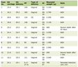 Click to view | Table 1. Clinical Timeline |
| Discussion | ▴Top |
Given the associated increased risk of prolonged hospital stay, in-hospital mortality, and overlapping features with other clinical syndromes, there is considerable importance placed on the identification and management of TCM [6]. Particularly in the peripartum period, TCM presents a diagnostic and management challenge, requiring a multidisciplinary approach to ensure the safety of both mother and child.
Differentiating TCM from other cardiomyopathies, particularly PPCM, demands careful consideration in order to understand prognosis and provide further counseling. A 2022 review elucidated distinguishing features of TCM in the postpartum period compared to PPCM (Table 2) [7–10]. TCM often presents more rapidly and over a shorter timeframe immediately after delivery in the setting of a clear emotional or physiological trigger, whereas PPCM develops more insidiously within the last few months of pregnancy and up to 6 months following delivery without a discrete precipitant.
Click to view | Table 2. Distinguishing Characteristics of Takotsubo CM and Peripartum CM |
In this case, the immediate temporal relationship with the emergency cesarean delivery and the onset of LV dysfunction strongly favored TCM. Data from the prospective nationwide RETAKO registry revealed that patients with peripartum-associated TCM had a higher prevalence of dyspnea, with atypical symptoms and ECG patterns including ST-segment elevation compared to TCM not associated with the peripartum period [8]. On TTE, TCM is most often characterized by regional wall motion abnormalities in a non-coronary distribution, typically with apical ballooning during systole and diastole, while PPCM displays global LV hypokinesis during systole and diastole [4, 7]. The pattern of hypokinesis of the basal-mid walls with apical sparing seen in this patient is consistent with the reverse TCM variant, which typically involves basal-mid wall hypokinesis to akinesis. This pattern did not fit the global dysfunction typical of PPCM [4]. Additionally, patients with TCM typically show a full recovery of LVEF within 1 month, whereas PPCM has a much more delayed LV function recovery [2, 4, 7]. Some studies have suggested that only 23–41% of patients with PPCM develop complete recovery of LVEF, whereas patients with TCM most often recover within days to weeks [7, 9, 10]. In our patient, normalization of LVEF from 30% to 51% within 3 days and 55–60% at 4 months is consistent with the expected recovery course of TCM and would be atypical for PPCM. Given the longer course for LV function recovery in PPCM, it carries higher rates of cardiovascular complications than TCM, including complex ventricular arrhythmias, chronic congestive heart failure, and mortality of 10–28% at 6 months and 2 years, respectively [7]. Accurate differentiation therefore has direct implications not only for prognosis but also for counseling regarding subsequent pregnancies.
ACS was also on the differential in this case, but, given the lack of ischemic changes in a territorial pattern on ECG and a modest troponin trend relative to the degree of dysfunction, this was felt to be less likely. Furthermore, wall motion patterns on echocardiography did not correspond to a single epicardial vessel distribution, as would typically be suggestive. Rapid recovery of LVEF also favored TCM. Thus, further evaluation of coronary anatomy via CT angiography or invasive coronary angiography was deferred.
Both TCM and PPCM pose difficulties in management, as most components of GDMT pose teratogenic risks. Angiotensin-converting enzyme (ACE) inhibitors and angiotensin II receptor blockers (ARBs), essential pharmacological treatments demonstrated to effectively reduce the recurrence of stress cardiomyopathy, are contraindicated in pregnancy due to an increased risk of major congenital cardiovascular anomalies, stillbirth, oligohydramnios, pulmonary hypoplasia, limb defects, fetal growth restriction, miscarriage, and preterm birth [4]. Other pillars of GDMT include mineralocorticoid receptor antagonists and sodium-glucose cotransporter 2 (SGLT2) inhibitors, which are also contraindicated during pregnancy due to concerns about feminization of male fetuses and renal dysfunction, respectively [11, 12]. Thus, peripartum treatment is focused on diuretics, beta blockers, afterload reduction with hydralazine or nitrates, and supportive care [11].
In the postpartum period, the addition of ACE inhibitors, ARBs, and mineralocorticoid receptor antagonists allows for more complete GDMT. For patients who are breastfeeding, medication selection should be guided by established lactation safety data. Enalapril is the preferred ACE inhibitor due to negligible breast milk levels, undetectable infant plasma levels, and absence of reported adverse effects in breastfed infants [12]. Metoprolol succinate is among the beta-blockers of choice during lactation, as only small amounts are excreted into breast milk, and no adverse reactions have been documented in breastfed infants [12]. Studies have shown that breastfeeding infants receive approximately 0.2% of maternal daily exposure to canrenone, the active metabolite of spironolactone, making it compatible with breastfeeding [12]. Furosemide and other diuretics are also generally safe for use during lactation [12]. For patients planning to breastfeed, counseling regarding the safety of medications should be conducted using evidence-based tools such as LactMed or in consultation with a maternal fetal medicine physician or obstetrician gynecologist. Cessation of lactation or “pump and dump” is not recommended for the large majority of patients.
Several studies have investigated the recurrence rate for TCM, with estimates varying widely, between 1% and 11.4% [13, 14]. In a study conducted by Santoro et al, there was no significant clinical evidence suggesting that the chronic management of TCM reduced the risk of recurrence [13]. Other studies have suggested that it is the trigger that affects prognosis, and that emotionally triggered TCM has more favorable short- and long-term prognosis compared to TCM secondary to neurologic disorders, physical activities, medical conditions, or procedures [15]. We hypothesize that, in our case, pain, anxiety, stress, and perhaps side effects of analgesia (as bupivacaine can cause hypertension in rare cases) led to hypertensive emergency in our patient [16, 17]. Due to impaired baroreceptor reflex function, which is known to be depressed during pregnancy, administration of labetalol and maternal hypotension led to a rapid swing in BP, leading to significant hypotension with impact on fetal health [16, 17]. Given the significant physiologic stress, including rapid fluctuations in BP and blood loss anemia, as well as emotional stress induced by this pregnancy, a catecholamine surge is suspected to have had direct myocardial stunning effects on beta adrenergic receptors [18]. Furthermore, labetalol blocks α1-, β1-, and β2-adrenoreceptors. The ratio of α- to β-blockade has been estimated to be approximately 1:7, which is greater when compared to other non-selective beta blockers such as carvedilol. This greater alpha-blocking effect may cause more hypotension, a significant concern in pregnant patients who may already be hemodynamically compromised [18].
Outcomes in subsequent pregnancies in patients with a history of peripartum TCM are not well known, likely due to the rarity of the condition. Nonetheless, thorough preconception counseling should be completed and include an evaluation of cardiac function with echocardiography. Subsequent monitoring throughout pregnancy should include serial echocardiography, N-terminal pro-brain natriuretic peptide monitoring, and continuation of beta-blocker therapy [19]. Timely initiation of contraception in postpartum individuals with TCM is critical in order to allow for recovery and prevention of short-interval pregnancy. Methods including long-acting reversible contraception (levonorgestrel or copper-containing intrauterine devices or the etonogestrel implant), progestin only oral contraceptives, and barrier methods are all safe for individuals who have cardiomyopathy. Per the United States Medical Eligibility Criteria (MEC) guidelines, combined oral contraceptives should be avoided in patients with cardiomyopathy, irrespective of time elapsed since the index event and the degree of cardiac dysfunction [20].
Although there are no specific guidelines for subsequent pregnancies after peripartum TCM, there are risk stratification guidelines based on LV function recovery that can be used to counsel these patients. The 2022 American Heart Association (AHA)/American College of Cardiology (ACC)/Heart Failure Society of America (HFSA) Heart Failure Guidelines highlight pre-pregnancy LVEF as the strongest predictor of outcomes in subsequent pregnancies [21]. Women with persistent LV dysfunction with LVEF less than 50% should be strongly discouraged from pregnancy due to increased risk of worsening LVEF and worse fetal outcomes [21]. Additional risk factor stratification and modification should be completed, including evaluation of other comorbidities such as hypertension, underlying anxiety or psychological stress, and diabetes. Discussions between the clinician and the patient should be transparent regarding the uncertain risk of recurrence and the potential for adverse fetal and maternal outcomes. Multidisciplinary management with maternal-fetal medicine, cardiology, and anesthesiology is essential should a patient have a subsequent pregnancy, in addition to close surveillance and echocardiographic monitoring.
Acknowledgments
This case was presented at the Congress on Cardiac Problems in Pregnancy in April 2025 in Vienna, Austria.
Financial Disclosure
The authors have no financial disclosures.
Conflict of Interest
The authors have no conflicts of interest to disclose.
Informed Consent
Not applicable, as there was no identifiable patient information.
Author Contributions
NT contributed to study design, literature search, manuscript drafting and editing. HW contributed to literature search, manuscript drafting and editing. KS contributed to literature review, manuscript drafting and editing. SB contributed to manuscript drafting and editing. AW contributed to manuscript drafting and editing. LK contributed to study design, literature search, manuscript drafting and editing.
Data Availability
The authors declare that data supporting the findings of this study are available within the article.
| References | ▴Top |
- Hurst RT, Prasad A, Askew JW, 3rd, Sengupta PP, Tajik AJ. Takotsubo cardiomyopathy: a unique cardiomyopathy with variable ventricular morphology. JACC Cardiovasc Imaging. 2010;3(6):641-649.
doi pubmed - Medina de Chazal H, Del Buono MG, Keyser-Marcus L, Ma L, Moeller FG, Berrocal D, Abbate A. Stress cardiomyopathy diagnosis and treatment: JACC State-of-the-Art review. J Am Coll Cardiol. 2018;72(16):1955-1971.
doi pubmed - Pelliccia F, Kaski JC, Crea F, Camici PG. Pathophysiology of takotsubo syndrome. Circulation. 2017;135(24):2426-2441.
doi pubmed - Yang WI, Moon JY, Shim M, Yang PS, Kang SH, Kim SH, Kim WJ, et al. Clinical features differentiating Takotsubo cardiomyopathy in the peripartum period from peripartum cardiomyopathy. Heart Vessels. 2020;35(5):665-671.
doi pubmed - Prasad A, Lerman A, Rihal CS. Apical ballooning syndrome (Tako-Tsubo or stress cardiomyopathy): a mimic of acute myocardial infarction. Am Heart J. 2008;155(3):408-417.
doi pubmed - Mogos MF, Ahn S, Muchira JM, Osmundson S, Piano MR. Pregnancy-associated takotsubo cardiomyopathy hospitalizations in the United States. Am J Physiol Heart Circ Physiol. 2023;325(3):H468-H474.
doi pubmed - Citro R, Radano I, Bellino M, Mauro C, Okura H, Bossone E, Akashy YJ. Epidemiology, pathogenesis, and clinical course of takotsubo syndrome. Heart Fail Clin. 2022;18(1):125-137.
doi pubmed - Vazirani R, Blanco-Ponce E, Almendro Delia M, Martin-Garcia AC, Fernandez-Cordon C, Uribarri A, Vedia O, et al. Peripartum takotsubo cardiomyopathy: a review and insights from a national registry. J Cardiovasc Dev Dis. 2024;11(2):37.
doi pubmed - Tzerefos S, Aloizou D, Nikolakopoulou S, Aloizos S. Takotsubo syndrome: differences between peripartum period and general population. Healthcare (Basel). 2024;12(16):1602.
doi pubmed - Garg H, Singh S, Ramachandran R, Trikha A. Takotsubo cardiomyopathy in pregnancy: A focused review. Journal of Obstetric Anaesthesia and Critical Care. 2023;13(2):142-159.
- Arany Z, Elkayam U. Peripartum cardiomyopathy. Circulation. 2016;133(14):1397-1409.
doi pubmed - Drugs and Lactation Database (LactMed®) [Internet]. Bethesda (MD): National Institute of Child Health and Human Development; 2006. Available from: https://www.ncbi.nlm.nih.gov/books/NBK501922/.
- Santoro F, Ieva R, Musaico F, Ferraretti A, Triggiani G, Tarantino N, Di Biase M, et al. Lack of efficacy of drug therapy in preventing takotsubo cardiomyopathy recurrence: a meta-analysis. Clin Cardiol. 2014;37(7):434-439.
doi pubmed - Almendro-Delia M, Lopez-Flores L, Uribarri A, Vedia O, Blanco-Ponce E, Lopez-Flores MDC, Rivas-Garcia AP, et al. Recovery of left ventricular function and long-term outcomes in patients with takotsubo syndrome. J Am Coll Cardiol. 2024;84(13):1163-1174.
doi pubmed - Ghadri JR, Wittstein IS, Prasad A, Sharkey S, Dote K, Akashi YJ, Cammann VL, et al. International expert consensus document on takotsubo syndrome (Part I): clinical characteristics, diagnostic criteria, and pathophysiology. Eur Heart J. 2018;39(22):2032-2046.
doi pubmed - Biaggioni I, Shibao CA, Diedrich A, Muldowney JAS, 3rd, Laffer CL, Jordan J. Blood Pressure management in afferent baroreflex failure: JACC review topic of the week. J Am Coll Cardiol. 2019;74(23):2939-2947.
doi pubmed - Lyon AR, Citro R, Schneider B, Morel O, Ghadri JR, Templin C, Omerovic E. Pathophysiology of takotsubo syndrome: JACC State-of-the-Art Review. J Am Coll Cardiol. 2021;77(7):902-921.
doi pubmed - Brooks VL, Cassaglia PA, Zhao D, Goldman RK. Baroreflex function in females: changes with the reproductive cycle and pregnancy. Gend Med. 2012;9(2):61-67.
doi pubmed - Park K, Bairey Merz CN, Bello NA, Davis M, Duvernoy C, Elgendy IY, Ferdinand KC, et al. Management of women with acquired cardiovascular disease from pre-conception through pregnancy and postpartum: JACC Focus Seminar 3/5. J Am Coll Cardiol. 2021;77(14):1799-1812.
doi pubmed - Centers for Disease Control and Prevention (CDC). Summary chart of U.S. medical eligibility criteria for contraceptive use (U.S. MEC). 2024. Accessed April 30, 2026.
- Heidenreich PA, Bozkurt B, Aguilar D, Allen LA, Byun JJ, Colvin MM, Deswal A, et al. 2022 AHA/ACC/HFSA guideline for the management of heart failure: a report of the American College of Cardiology/American Heart Association Joint Committee on Clinical Practice Guidelines. Circulation. 2022;145(18):e895-e1032.
doi pubmed
This article is distributed under the terms of the Creative Commons Attribution 4.0 International License (CC BY 4.0), which permits unrestricted use, distribution, and reproduction in any medium, including commercial use, provided the original work is properly cited.
Journal of Medical Cases is published by Elmer Press Inc.