| Journal of Medical Cases, ISSN 1923-4155 print, 1923-4163 online, Open Access |
| Article copyright, the authors; Journal compilation copyright, J Med Cases and Elmer Press Inc |
| Journal website https://jmc.elmerpub.com |
Case Report
Volume 17, Number 9, September 2026, pages 529-536

Catastrophic Outcome of Pregnancy Complicated With Tricuspid Valve Nonbacterial Thrombotic Endocarditis
Jake Smitha, Juan Pinillab  , Ricardo F.O. Suruagy Mottac, Chiranjiv Virka, Kalgi Modia, d
aDepartment of Cardiovascular Medicine, Louisiana State University Health, New Orleans, LA, USA
bCES University School of Medicine, Medellin, Colombia
cDepartment of Medicine, Cesmac University Center, Maceio, Alagoas, Brazil
dCorresponding Author: Kalgi Modi, Department of Cardiovascular Medicine, Louisiana State University Health, New Orleans, LA, USA
Manuscript submitted February 2, 2026, accepted April 15, 2026, published online July 28, 2026
Short title: Fatal Tricuspid NBTE Complicating Pregnancy
doi: https://doi.org/10.14740/jmc5270
| Abstract | ▴Top |
Nonbacterial thrombotic endocarditis (NBTE) is a rare condition characterized by sterile fibrin-platelet vegetations on cardiac valves, typically associated with hypercoagulable states. Isolated tricuspid valve (TV) involvement during pregnancy is exceedingly rare, with no previously reported cases of fatal maternal and fetal outcome in the published literature. We report a 31-year-old woman (gravida 3) with a complex medical history including human immunodeficiency virus infection, chronic kidney disease, recurrent venous thromboembolism, and thrombocytopenia, who presented at 9 weeks of gestation with a large, mobile TV mass. Initial workup revealed elevated IgM antiphospholipid antibodies, a positive dilute Russell’s viper venom time, negative blood cultures, and a concurrent pulmonary embolism. A transthoracic echocardiogram identified a 2.2 × 1.6 cm irregular hyperechoic mass on the anterior tricuspid leaflet. Despite therapeutic anticoagulation with enoxaparin and aspirin, the mass enlarged to 3.5 × 2.7 cm by 13 weeks of gestation. Following multidisciplinary discussion and shared decision-making with the patient, AngioVac-assisted percutaneous aspiration thrombectomy was performed at 14 weeks and 4 days of gestation. Histopathology confirmed sterile fibrin thrombi with histiocytic infiltration consistent with NBTE. Fetal demise was identified on postoperative day 2. Subsequently, the patient developed encephalopathy and massive hemoptysis, raising concern for catastrophic antiphospholipid syndrome. Despite aggressive resuscitation including venovenous extracorporeal membrane oxygenation, continuous renal replacement therapy, and maximum vasopressor support, the patient died. This is the first reported case of fatal maternal and fetal outcomes associated with TV NBTE during pregnancy. The case underscores the challenges of evaluation and management of cardiac masses in immunocompromised pregnant patients, the limitations of APS classification criteria in the peripartum setting, and the importance of a multidisciplinary cardio-obstetric approach.
Keywords: Endocarditis; Thrombus; Tricuspid valve
| Introduction | ▴Top |
Nonbacterial thrombotic endocarditis (NBTE) is a rare condition characterized by the formation of sterile fibrin-platelet vegetations on the surface of cardiac valve leaflets, most commonly the mitral or aortic valves. Tricuspid valve (TV) involvement is uncommon and represents a particularly challenging clinical entity. NBTE typically occurs in association with hypercoagulable states, malignancies, or autoimmune disorders—most notably antiphospholipid syndrome (APS) and systemic lupus erythematosus. The most frequent clinical presentation is an embolic event, with cerebral emboli occurring more commonly than peripheral thromboembolic complications [1].
Contemporary data from large tertiary care centers suggest that NBTE is diagnosed more frequently in females. Its occurrence during pregnancy is exceedingly rare, and the true epidemiology remains poorly defined due to the difficulty of premortem diagnosis and the scarcity of reported cases in the literature [2]. Pregnancy itself constitutes a physiological prothrombotic state, driven by increases in clotting factors, decreased protein S activity, and reduced fibrinolysis. When superimposed on additional risk factors such as HIV infection, chronic kidney disease, recurrent venous thromboembolism, and antiphospholipid antibodies, this prothrombotic milieu may become severe enough to precipitate catastrophic cardiovascular and obstetric complications.
The AngioVac system is a percutaneous aspiration thrombectomy device that offers a minimally invasive alternative to open cardiac surgery for the debulking of right-sided intracardiac masses, including vegetations associated with NBTE. The device utilizes venous drainage and return cannulae connected to an extracorporeal circuit with a pump and filter, allowing thrombotic material to be aspirated, filtered, and reinfused while minimizing hemodynamic compromise [3]. Its use during pregnancy remains anecdotal, with only a small number of cases reported in the literature.
We present what is, to our knowledge, the first reported case of TV NBTE managed with AngioVac-assisted percutaneous aspiration thrombectomy during pregnancy, resulting in both maternal and fetal fatality. This case highlights the diagnostic challenges, therapeutic dilemmas, and catastrophic potential of this condition in the peripartum setting, and underscores the critical value of a multidisciplinary cardio-obstetric approach in managing high-risk thrombotic disease during gestation.
| Case Report | ▴Top |
History of presentation
A 31-year-old G3P1011 Black female, 4 months prior to presentation, had been diagnosed with a TV mass at facility out-of-state. Blood cultures obtained were negative; however, infectious diseases recommended an empiric 12 weeks of intravenous antibiotics for culture negative endocarditis. She was also initiated on warfarin. She presented to our facility with a chief complaint of shortness of breath and chest pain. Initial vital signs showed the patient to be afebrile; however, she was hypotensive, tachycardic, and dyspneic with oxygen saturation of 100% on room air. Her heart exam was regular without murmur, lung exam was clear to auscultation, extremities without edema.
Past medical history
Her past medical history included human immunodeficiency virus (HIV), miscarriage, hypertension, thrombocytopenia, recurrent venous thromboembolism (splenic thrombus, deep vein thrombosis, and pulmonary embolism since her second pregnancy in 2022 with a reported apixaban failure and on warfarin) and chronic kidney disease.
Differential diagnosis
The patient’s presentation may be explained by several alternative etiologies, including culture-negative infective endocarditis with systemic sepsis, thromboembolic phenomena such as pulmonary embolism, or hemodynamic deterioration secondary to a large obstructive intracardiac mass in the setting of a severe prothrombotic state.
Investigation
Laboratory workup was significant for a hemoglobin 9.8 g/dL, hematocrit 31%, platelet count 50 × 103/µL, creatinine 2.4 mg/dL, erythrocyte sedimentation rate 120 mm/h, and a rheumatoid factor 25 IU/mL. Importantly, beta-2 GP1 IgM antibody 20.6 (ref < 15), markedly elevated phospholipid antibody IgM level of 142.6 MPL (reference < 15 MPL), dilute Russell’s viper venom time (DRVVT) ratio was 1.99 (normal < 1.2), negative IgG phospholipid antibody, and lupus anticoagulants. Her international normalized ratio (INR) was 1.1. A computed tomography (CT) pulmonary angiogram confirmed the presence of a pulmonary embolism in the right lower lobe without evidence of right heart strain. Despite intrauterine device in situ, she was found to be pregnant. Obstetrics was consulted who performed a transvaginal ultrasound showing a live intrauterine pregnancy of approximately 9 weeks as well as a small chorionic hemorrhage. Transthoracic echocardiography revealed a large, mobile, irregular, hyperechoic mass measuring 2.2 × 1.6 cm attached to the anterior leaflets of the TV, moderate tricuspid regurgitation, and normal biventricular size and function (Supplementary Video 1, jmc.elmerpub.com).
Management
Multiple disciplines were arranged including cardiology, cardiothoracic surgery, vascular surgery, maternal and fetal medicine, hematology, rheumatology, infectious disease, and nephrology. Cardiothoracic surgery recommended no acute surgical intervention due to high risk of fetal demise but to consider aspiration thrombectomy once the fetus is in the second trimester. Maternal and fetal medicine recommended prioritization of the mother due to the concern that uncontrolled cardiac maladies posed the greatest threat to fetal demise. Infectious diseases recommended against further antibiotics as multiple sets of blood cultures were negative. After discussion, she opted not to terminate the pregnancy and was treated with aspirin and anti-Xa-level-guided enoxaparin.
She presented at 13 weeks of gestation with worsening chest pain and dyspnea; follow-up echocardiography showed interval growth of the mass despite adequate anticoagulation (1.07 anti-Xa units/mL). A transesophageal echocardiogram revealed a larger, hypermobile, irregular “fleur-de-lis” shaped tricuspid mass measuring 3.5 × 2.7 cm attached to anterior leaflet with prolapse into the right ventricle during diastole (Supplementary Video 2, jmc.elmerpub.com, Fig. 1). With a multidisciplinary approach along with shared decision-making with the patient, the decision was made to perform aspiration thrombectomy. The patient was bridged with heparin and underwent transesophageal echocardiogram–guided AngioVac-assisted percutaneous aspiration thrombectomy at 14 weeks 4 days gestation under general anesthesia (Fig. 2). The fetus was shielded with eight layers of 0.5 mm lead apron, with a total 8.8 min of fluoroscopy time at exposure rate of 3 frames per second (FSP) with lowest pulse rate, and total 120 milligray (mGy). The procedure resulted in debulking of the mass to 1.5 cm. The patient tolerated the procedure well, and fetal heart tones before and after the procedure were appropriate. Pathology of the tricuspid mass showed a sterile, fibrin thromboembolism surrounded by histiocytes (Figs. 3–6). She was transitioned to therapeutic enoxaparin dose per anti-Xa levels.
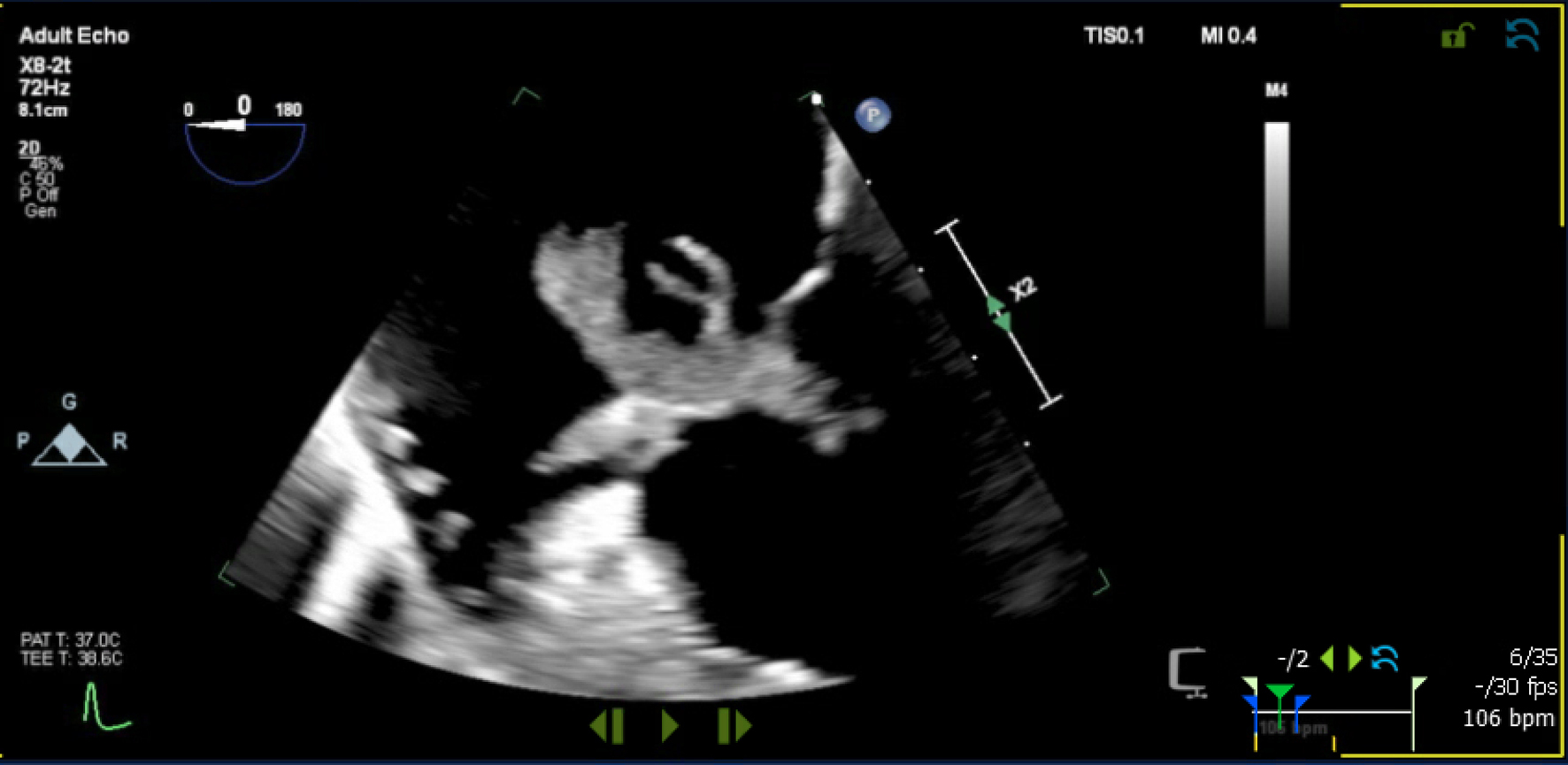 Click for large image | Figure 1. Transesophageal image showing a large multi-lobed mass attached to anterior leaflet of tricuspid valve. |
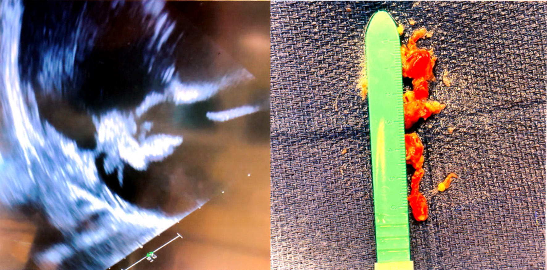 Click for large image | Figure 2. Aspirated mass from tricuspid valve with AngioVac thrombectomy catheter. |
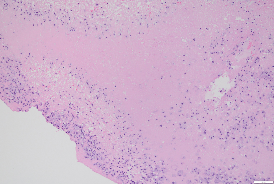 Click for large image | Figure 3. A fibrin thrombus is observed, surrounded by a dense accumulation of histiocytes. |
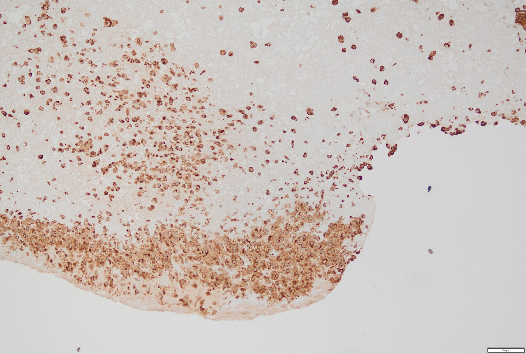 Click for large image | Figure 4. Immunohistochemical staining for CD68 highlights the histiocytes. |
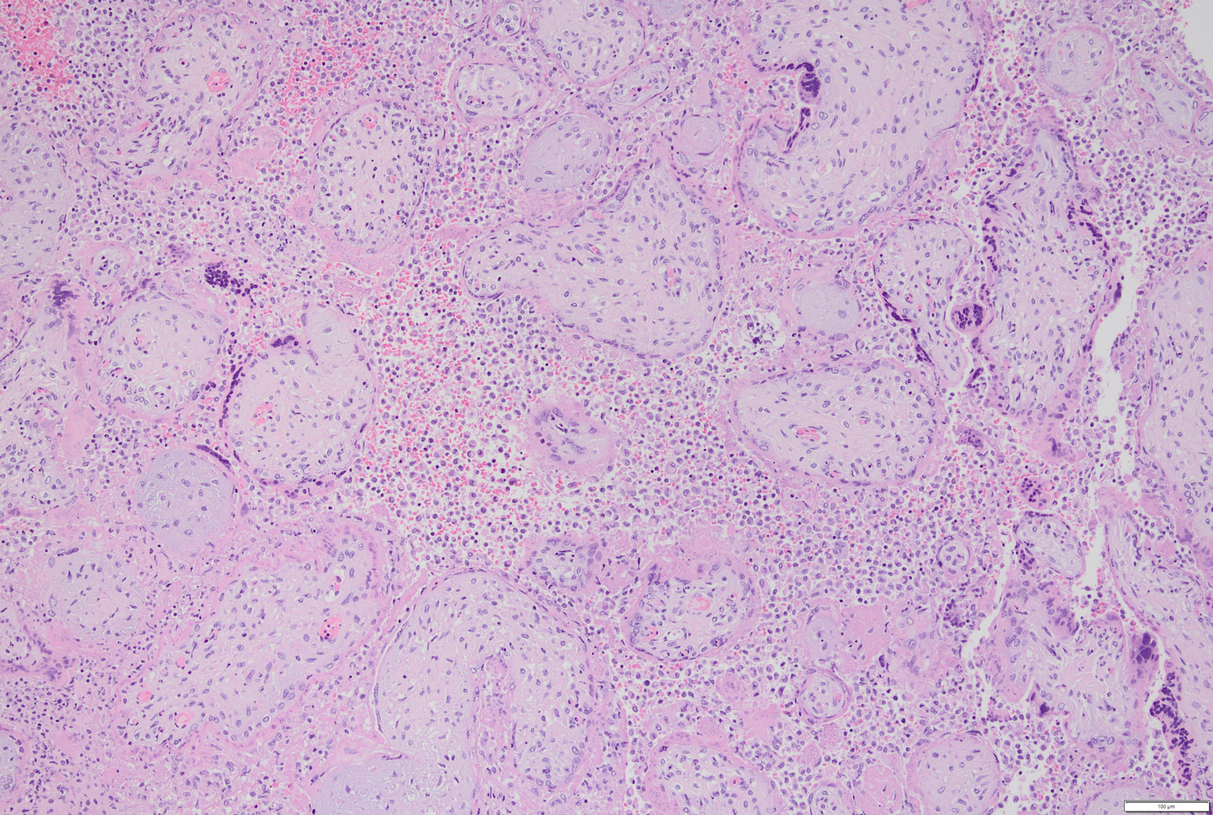 Click for large image | Figure 5. The intervillous space is filled with sheets of histiocytes. |
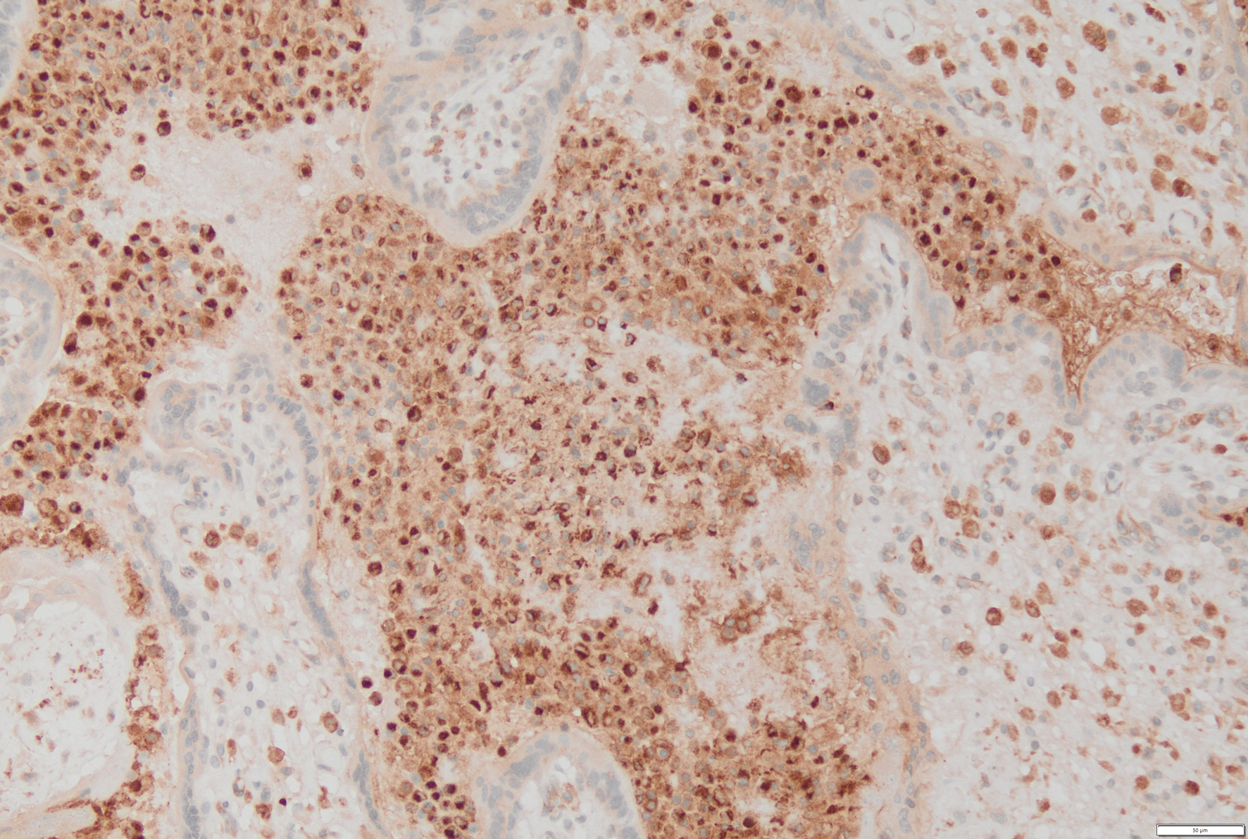 Click for large image | Figure 6. CD68 immunostaining further confirms the presence of histiocytes in the intervillous space. |
Outcome
On post-operative day 2, a routine check showed no fetal heart tones, unfortunately confirming fetal demise just shy of 15 weeks gestation. Interestingly pathology of the placenta shared similar findings to TV mass with chronic histiocytic intervillositis with increased perivillous fibrin deposition. Five days after discharge, she again returned with sharp chest pain. Workup including electrocardiogram (EKG), troponins, and chest CT were all unremarkable. Repeat echo was without right ventricle (RV) strain and a decreased tricuspid mass size. During this hospitalization, her mentation became altered. The differential diagnoses included sepsis from the right groin access site, retained vaginal products, central nervous system (CNS) infection, and heparin-induced thrombocytopenia. She was treated with intravenous immunoglobulin (IVIG) and steroids. During hospitalization, she developed massive hemoptysis with bronchoscopy showing right upper lobe bronchus bleeding with pooling into her right lower lobe bronchus. Her condition worsened and despite aggressive resuscitation efforts which included a trial of venovenous extracorporeal membrane oxygenation (VV ECMO), continuous renal replacement therapy (CRRT), and maximum pressor support, the patient succumbed (Fig. 7).
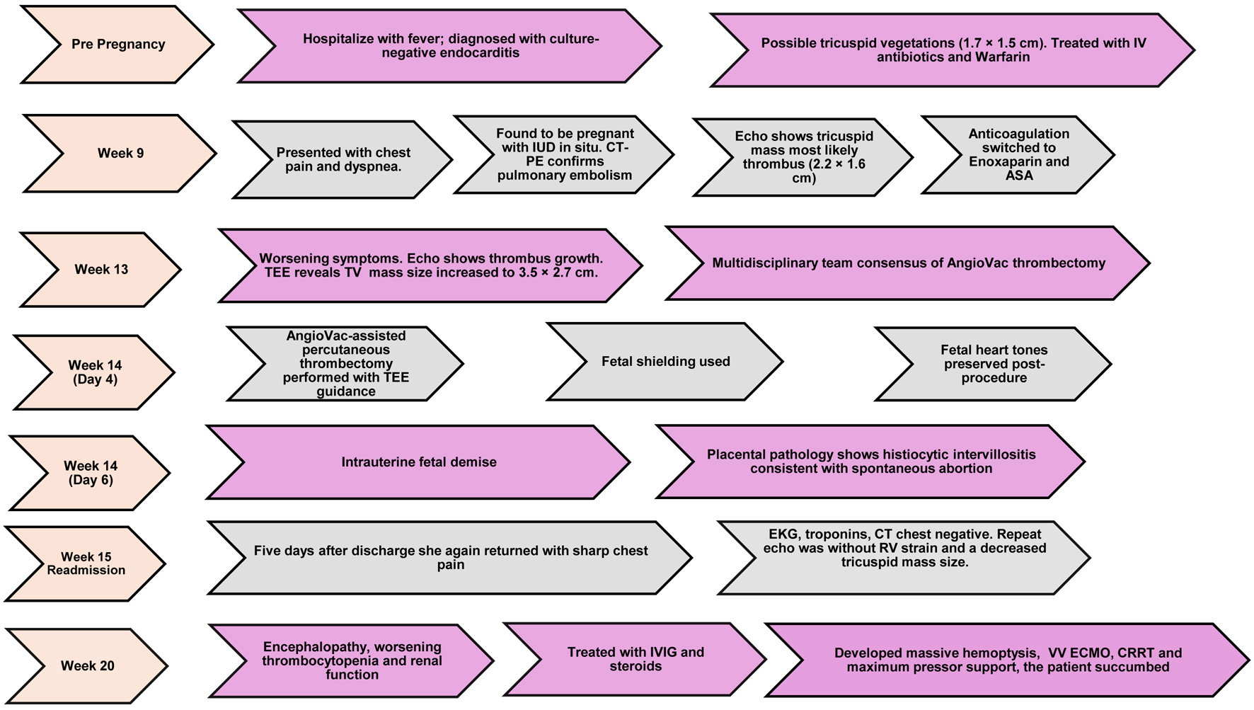 Click for large image | Figure 7. Central illustration capturing the clinical course. |
| Discussion | ▴Top |
This case of a pregnant woman with a large TV mass, whose peripartum course was complicated by both maternal and fetal catastrophe, raises several clinical points.
First, our case illustrates the clinical constellation of suspected APS, with thrombi involving the placenta, lung, and TV, alveolar hemorrhage, and encephalopathy. It is important to acknowledge, however, that our patient did not fulfill the updated international classification criteria for definite APS, as her IgG anticardiolipin antibody was negative, lupus anticoagulant was negative, and repeat confirmatory testing at 12 weeks was not possible given her clinical deterioration [4]. The sole positive finding was a high-titer IgM anticardiolipin antibody. Notably, transient or false-positive IgM antiphospholipid antibodies are well-documented in HIV-infected patients, attributable to polyclonal B-cell activation rather than true autoimmune pathology [5]. This represents an important confounding factor in our case, and the IgM positivity should therefore be interpreted with caution. Nevertheless, IgM anticardiolipin antibodies are present in up to 82% of pregnancies complicated by APS and carry substantial predictive value for obstetric and thrombotic events [6]. Furthermore, cases of catastrophic APS with isolated IgM anticardiolipin positivity in the absence of IgG antibodies have been reported [7], and it is possible that the diffuse coagulopathy observed in our patient was triggered by the invasive thrombectomy procedure in the setting of a pre-existing prothrombotic state. Our case highlights the importance of screening for APS in patients with complex obstetric and thrombotic histories, and supports reconsidering isolated IgM-positive suspected catastrophic APS as a contributor to peripartum morbidity.
An important consideration in this case is the exclusion of occult or culture-negative infectious endocarditis, particularly given our patient’s severely immunocompromised state secondary to HIV infection and chronic kidney disease. Although multiple sets of blood cultures obtained throughout her clinical course remained negative, and infectious diseases consultants recommended against further antibiotic therapy, we acknowledge that additional microbiological data would have strengthened the case for NBTE over culture-negative infectious endocarditis. Patient had a normal absolute CD4 lymphocyte count of 550 and HIV viral load of < 20 copies/mL at the time of presentation, as well as negative screening for opportunistic infections including tuberculosis, and endemic fungi from blood and negative aerobic, anaerobic, mycobacteria, fungal culture, and KOH prep from heart valve tissue. Several key features support a diagnosis of NBTE rather than infectious endocarditis: (1) the absence of fever and systemic signs of infection; (2) the atypical echocardiographic morphology of the mass, characterized by an irregular, hypermobile, multi-lobed appearance without valve leaflet destruction to cause significant tricuspid regurgitation is distinct from the typical infectious endocarditis; and (3) histopathological confirmation of sterile fibrin thrombus without evidence of microorganisms or acute inflammatory infiltrate [2].
Second, the evaluation of a cardiac mass during pregnancy presents substantial diagnostic challenges. Given our patient’s negative blood cultures and the atypical echocardiographic features inconsistent with location for primary cardiac tumors, absence of significant tricuspid regurgitation, and attachment to hypermobile structure, a nonbacterial thrombotic sterile vegetation was highest on our differential. Our patient harbored multiple predisposing factors including HIV infection, chronic kidney disease, recurrent venous thromboembolism, pregnancy, and possible APS, all of which may have synergistically contributed to the development of NBTE. It is important to note that in the setting of severe systemic inflammation, multifactorial hypercoagulability, and recurrent thromboembolism, an organized intracardiac thrombus arising from the patient’s complex prothrombotic state represents a viable alternative explanation. The histopathological distinction between primary NBTE and secondary organized thrombus can be subtle, as both may demonstrate sterile fibrin deposition without organisms or acute inflammation. Primary NBTE is characterized by bland, avascular fibrin-platelet deposits with minimal inflammatory infiltrate on the valve surface, without neovascularization or granulation tissue. In contrast, an organized thrombus demonstrates progressive fibroblastic ingrowth, neovascularization, and heterogeneous inflammatory infiltrate over time. In our case, the pathological findings—sterile fibrin thrombus with histiocytic infiltration rather than fibroblastic organization, mirrored by similar fibrinous pathology in the placenta—combined with the absence of organisms and the clinical context of systemic hypercoagulability, favor primary NBTE or an autoimmune-driven fibrinous process over simple organized thrombus [8]. We acknowledge, however, that definitive differentiation between these entities in the setting of severe systemic illness remains challenging and that both mechanisms may have contributed (Fig. 8).
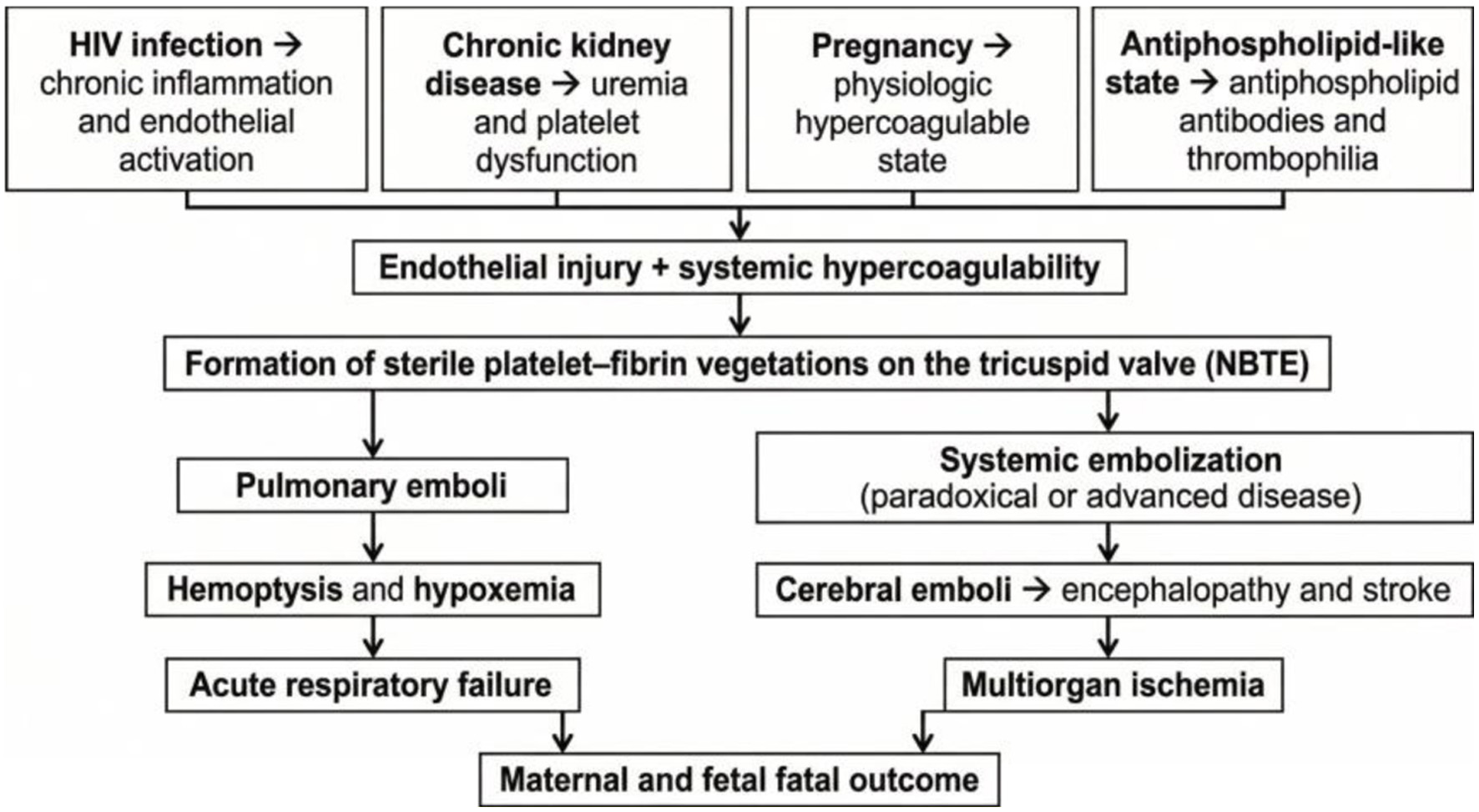 Click for large image | Figure 8. Proposed pathophysiological mechanism of tricuspid valve nonbacterial thrombotic endocarditis (NBTE) during pregnancy. |
Third, isolated tricuspid NBTE is quite rare, thus the optimal management of this condition in pregnancy is anecdotal. Despite therapeutic anticoagulation, our patient experienced symptomatic thrombus enlargement. Up to 30% of patients with confirmed APS treated with low molecular weight heparin and aspirin continue to have maternal fetal complications [9]. Given the risk of embolization and hemodynamic compromise, the team proceeded with AngioVac-assisted percutaneous thrombus removal, as open surgery was considered prohibitively high-risk [10]. The AngioVac system is a minimally invasive aspiration thrombectomy device with increasing evidence supporting its use in right-sided cardiac thrombi, including rare cases during pregnancy. Three previously published case reports described successful AngioVac-assisted thrombus removal followed by cesarean delivery in patients with right-sided endocarditis with favorable maternal and fetal outcomes. In a meta-analysis, the pooled event rate for procedure success was 80.5 (CI: 70.0–88.0) and mortality was 14.8 (CI: 8.5–24.5) among right atrial/caval thrombi cases. These patients generally exhibited fewer comorbidities and greater physiologic reserve compared with our patient [3, 11, 12]. Similarly, a surgically managed case of Libman-Sacks endocarditis associated with APS and subsequent successful delivery involved a patient with minimal additional comorbidities [10].
Although catastrophic APS remains the leading diagnosis, alternative explanations for the post-procedural deterioration merit consideration. Pulmonary hemorrhage may have resulted from anticoagulation, uremic coagulopathy, extracorporeal circuit–induced inflammation, or HIV-related vasculopathy. Encephalopathy could be explained by septic embolization from the groin access site or retained products of conception, providing a unifying mechanism for both neurological decline and massive hemoptysis. The absence of a post-mortem examination precludes definitive attribution of the terminal event.
Fourth, from a pathological standpoint, the size of the TV NBTE in our patient represents one of the largest reported to date. Notably, both the valve mass and placenta demonstrated similar histiocytic inflammatory infiltrates, suggesting an autoimmune mechanism and a potential role for immunomodulatory therapy to prevent clinical events.
Fifth, from an ethical perspective, this case illustrates the complexities of decision-making in high-risk pregnancies. The patient elected to continue her pregnancy despite significant maternal and fetal risks. The multidisciplinary approach facilitated informed, patient-centered decision-making that balanced maternal autonomy, procedural risks, and fetal considerations. Although thrombus removal was technically successful and fetal stability was temporarily achieved, intrauterine fetal demise and maternal mortality occurred later, likely related to the patient’s extensive comorbidities and persistent pro-thrombotic state.
Conclusion
This is the first reported case of fatal maternal and fetal outcomes associated with TV NBTE during pregnancy managed with AngioVac-assisted percutaneous aspiration thrombectomy. This case underscores the diagnostic challenges of evaluating cardiac masses in severely immunocompromised pregnant patients and highlights the limitations of current APS classification criteria in the peripartum setting. NBTE should be considered in pregnant patients with unexplained cardiac masses. Suspected catastrophic APS with isolated IgM positivity should remain in the differential diagnosis of peripartum multi-organ failure even when formal classification criteria are not met. Multidisciplinary cardio-obstetric management is essential when anticoagulation alone is insufficient.
| Supplementary Material | ▴Top |
Suppl 1. Transthoracic echocardiogram 2D, right ventricular inflow view showing large, irregular, multi lobed, and mobile mass attached to the atrial aspect of the anterior leaflet of Tricuspid valve.
Suppl 2. Transesophageal echocardiogram 2D, lower esophageal view showing “fleur-de-lis” shaped large, irregular, multi lobed, and mobile mass attached to the anterior leaflet of Tricuspid valve.
Acknowledgments
None to declare.
Financial Disclosure
None to declare.
Conflict of Interest
The authors have no relevant financial or non-financial interests to disclose. This research did not receive any specific grant from funding agencies in the public, commercial, or not-for-profit sectors.
Informed Consent
Informed consent has been obtained.
Author Contributions
Jake Smith, MD, collected and curated the clinical data, verified the clinical accuracy of the case, finalized the manuscript, and serves as the first author. Juan Pinilla performed the initial literature review and drafted the first version of the manuscript. Ricardo F.O. Suruagy Motta contributed to the preparation of figures and illustrations and assisted with visual presentation of the case. Chiranjiv Virk, MD, and Kalgi Modi, MD, provided senior clinical supervision, critically reviewed the manuscript for important intellectual content, and guided the final revisions. All authors reviewed and approved the final version of the manuscript and agreed to be accountable for all aspects of the work.
Data Availability
The authors declare that data supporting the findings of this study are available within the article.
Abbreviations
APS: antiphospholipid syndrome; CNS: central nervous system; CRRT: continuous renal replacement therapy; CT: computed tomography; DRVVT: dilute Russell’s viper venom time; ECMO: extracorporeal membrane oxygenation; EKG: electrocardiogram; ESR: erythrocyte sedimentation rate; FSP: frames per second; G3P0010: gravida 3, para 0, abortus 1, living 0; HIV: human immunodeficiency virus; INR: international normalized ratio; IVIG: intravenous immunoglobulin; mGy: milligray; NBTE: nonbacterial thrombotic endocarditis; RV: right ventricle; TEE: transesophageal echocardiogram; TTE: transthoracic echocardiogram; TV: tricuspid valve; VV ECMO: venovenous extracorporeal membrane oxygenation
| References | ▴Top |
- Ahmed O, King NE, Qureshi MA, Choudhry AA, Osama M, Zehner C, Ali A, et al. Non-bacterial thrombotic endocarditis: a clinical and pathophysiological reappraisal. Eur Heart J. 2025;46(3):236-249.
doi pubmed - Quintero-Martinez JA, Hindy JR, El Zein S, Michelena HI, Nkomo VT, DeSimone DC, Baddour LM. Contemporary demographics, diagnostics and outcomes in non-bacterial thrombotic endocarditis. Heart. 2022;108:1637-1643.
doi pubmed - Hameed I, Lau C, Khan FM, Wingo M, Rahouma M, Leonard JR, Di Franco A, et al. AngioVac for extraction of venous thromboses and endocardial vegetations: a meta-analysis. J Card Surg. 2019;34(4):170-180.
doi pubmed - Miyakis S, Lockshin MD, Atsumi T, Branch DW, Brey RL, Cervera R, Derksen RH, et al. International consensus statement on an update of the classification criteria for definite antiphospholipid syndrome (APS). J Thromb Haemost. 2006;4(2):295-306.
doi pubmed - Petrovas C, Vlachoyiannopoulos PG, Kordossis T, Moutsopoulos HM. Anti-phospholipid antibodies in HIV infection and SLE with or without anti-phospholipid syndrome: comparisons of phospholipid specificity, avidity and reactivity with beta2-GPI. J Autoimmun. 1999;13(3):347-355.
doi pubmed - Yang Y, Jiang H, Tang Z, Pan H, Liu H, Cheng X, Su Y, et al. Assessment of the 2023 ACR/EULAR antiphospholipid syndrome classification criteria in a Chinese cohort: Impact on clinical practice. J Autoimmun. 2024;146:103237.
doi pubmed - Kennedy M, Jackson J, Khan I, Keeling PW, Gaffney E. Catastrophic anti-phospholipid syndrome in the absence of IgG anti-cardiolipin antibodies. Scand J Rheumatol. 1995;24(6):389-391.
doi pubmed - Eiken PW, Edwards WD, Tazelaar HD, McBane RD, Zehr KJ. Surgical pathology of nonbacterial thrombotic endocarditis in 30 patients, 1985-2000. Mayo Clin Proc. 2001;76(12):1204-1212.
doi pubmed - Radin M, Cecchi I, Schreiber K, Rubini E, Roccatello D, Cuadrado MJ, Sciascia S. Pregnancy success rate and response to heparins and/or aspirin differ in women with antiphospholipid antibodies according to their Global AntiphosPholipid Syndrome Score. Semin Arthritis Rheum. 2020;50(3):553-556.
doi pubmed - Migliorini S, Santoro C, Scatteia A, Dellegrottaglie S, Tufano A, Cuomo V, Pilato E, et al. A rare case of tricuspid valve libman-sacks endocarditis in a pregnant woman with primary antiphospholipid syndrome. J Clin Med. 2022;11(19):5875.
doi pubmed - Ayzenbart V, Fuentes H, Fuentes F, Aziz S, Joseph M. Angiovac use in endocarditis during pregnancy: a novel approach for recurrent debulking of tricuspid infective vegetations in a 27-year-old woman in her 22nd and 26th weeks of pregnancy. Chest. 2021;160:A786.
doi - Boudova S, Casciani T, Weida J. Percutaneous debulking of tricuspid vegetations due to infectious endocarditis in pregnancy: a case report. AJOG Glob Rep. 2023;3(2):100204.
doi pubmed
This article is distributed under the terms of the Creative Commons Attribution 4.0 International License (CC BY 4.0), which permits unrestricted use, distribution, and reproduction in any medium, including commercial use, provided the original work is properly cited.
Journal of Medical Cases is published by Elmer Press Inc.